A cell is the structural and functional unit of life. It is the building block of which all living organisms are made, and the smallest unit of life capable of all the living functions. It is defined as a mass of protoplasm bounded by a plasma membrane.
Cell vary in size. Most cells are very small (microscopic), some may be very large. Some cells may exist as independent units of life. Some such cells like Euglena and Amoeba can change their shape, but most cells have a fixed shape. The number of cells vary from organism to organism. An amoeba is single-celled, while a human body weighing about 60 kg may have as many as 60 × 1015 cells. In unicelluar organisms, e.g. Amoeba, Paramoecium or Chalamydomonas, all the basic functions of a living being are performed in one cell, while multicellular organisms have well-developed division of labour. So, their different functions are performed by different organs. For example, we have a stomach to digest food, a heart to pump blood and a brain to think.
An English scientist, Robert Hooke, discovered the cell in 1665 while examining thin sections of cork under his simple microscope. He observed a mass of hexagonal chambers like a honeycomb and called them(compartments) cells. Cell is the Latin word for 'a little room'.
DISCOVERY OF CELL
1. Robert Hooke (1665) :– An English man and first 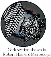
curator of Royal society of London.
Observed a thin transverse section of bark of a
tree under self designed microscope.
He noticed honey - comb like compartments.
He coined the term cell.
He wrote a book - Micrographia.
He actually observed dead cells.
2. Antony Van Leeuwenhoek (1674) was first to observe
living cells like bacteria [from tartar of teeth]
erythrocytes [fish], sperms and protozoans [eg. Vorticella]
3. N. Grew (1682) :– Proposed cell concept which states that cell is unit of structure of organisms.
4. Rudolf Virchow (1858) :– Proposed that new cells formed from the pre-existing cells.
MICROSCOPE
A microscope is an instrument to view small objects by magnifying them. It enables us to see the different types of living cells and the structures they contain.

Types of microscopes
There are mainly three types of microscopes. They are :
1. Light microscope : The light microscope uses light to produce images.
2.Transmission Electron Microscope (TEM) : The electron microscope was designed by Knoll & Ruska (1932). A TEM makes use of a beam of highly energetic electrons to examine objects. The image produced is of a very fine scale.
3. Scanning Electron Microscope (SEM) : Like the TEM, the SEM also uses electrons to produce images. In the case of a SEM, electrons are reflected off the surface of the specimen, because of which SEM images usually manage to capture the physical features of a cell in great detail.
STRUCTURE OF A CELL
* Cell membrane or plasma membrane:
A cell is essentially a tiny 'bag' of living matter. The covering of this 'bag' is called the cell membrane or plasma membrane. It maintains the shape and size of the cell and protects its contents. It acts like a sentry-allowing only some things to enter and leave the cell and stopping others. For example, it allows oxygen and nutrients to pass into the cell and lets wastes pass out of it. This is why it is called selectively permeable.
* Cell wall: Plant cells have an additional protective wll called the cell wall. It is thick, rigid and permeable, and is made up of a carbohydrate called cellulose.
* Cytoplasm :The matter inside the cell mebrane is called cytoplasm. It consists of a jellylike fluid with various structures, such as the nucleus, floating in it. These structures are called organelles.
Salts, proteins, sugar and other substances are dissolved in the fluid.
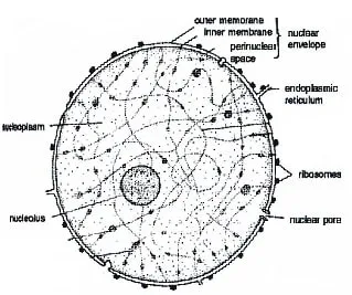
* Nucleus:Almost every cell has a nucleus. Red blood cells are among the exceptions. The nucleus is the largest and the most important organelle of the cell. It is usually spherical or oval in shape. Inside it there are thread like structures called chromosomes. Nucleus is the controlling central cell. Chromosomes have genes arranged in a linear fashion.
It is the most important part of the living cell.
It is usually spherical or oval in shape.
It controls all the vital functions of the cell.
It has four components:
(i) Nulcear Membrane (ii) Nulceoplasm
(iii) Nulceolus (iv) Chromosomes
(i) Nuclear membrane: Surrounds the nucleus and separates it from the cytoplasm. It is permeable and controls the passage of materials between cytoplasm and nucleoplasm
(ii) Nucleoplasm: The part of protoplasm which is enclosed by nuclear membrane is called nucleoplasm. It contains chromatin threads and nucleolus.
(iii) Nulceolus: It is a spherical body in the nucleus. It is composed of RNA and is responsible for protein synthesis.
(iv) Chromosomes: Nucleus contains thread like structures called chromosomes.
The hereditary units of chromosomes are the genes. They are responsible for the transmission of characters from parents to the offsping.
Nucleus along with tis role in inheritance regulates and controls differeny metabolic activities of the cell.
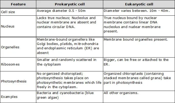
On the basis of well organised nucleus, cells can be of two types
(i) Prokaryotic cell (ii) Eukaryotic cell
(i) Prokaryotic cells: These are cells having primitive nucleus without nuclear membrane. Organism with primitive nucleus are known as prokaryotes.
Ex. Bacteria and blue green algae.
(ii) Eukaryotic cells: These are cells having a well organised nucleus with nuclear membrane. Organsims with true nucleus are known as eukaryotes.
Ex. Man, elephant, onion.
* Vacuoles:
The central part of most plant cells is occupied by a large vacuole. You may have noticed it in some of the plant cells you observed. It is a sac like structure filled with fluid. Food, wastes pigments and other substances are dissolved in the fluid. Some plant cells have a number of large vacuoles.
Vaculoes are not so common in animal cells. When they ocour, they are much smaller in size.
* Plastids:
These organelles are not present in animal cells. Chloroplasts (a type of plastid) contain the green pigment chlorophyll and are responsible for photosynthesis. Only green parts of plants have chloroplasts.
There are two other types of plastids called chromoplasts and leucoplasts. Chromoplasts contain pigments which give fruits and flowers their colours. Leucoplasts store food and are found in the storage organs of plants.
* Endoplasmic reticulum (ER)
The endoplasmic reticulum is a network of tube-like structures running through the cytoplasm. If ribosomes are attached to it, the reticulm is rough, otherwise it is smooth.
Function - It gives internal support to the colloidal matrix (cytoplasm).
Rough endoplamic reticulum (RER) is associated with the synthesis of proteins.
* Ribosomes
Ribosomes are extremely amall, round bodies found either in the state in the cytoplasm or attached to the surface of the ER. They are composed of ribonucleoprotein (ribonucleic acid and protein).
Functions - The main function of ribosomes is to act as a platform or work place for the synthesis of proteins.
* Mitochondria
Mitochondria are small, rod shaped organelles found in large numbers. Each mitochondrion is bounded by two membranes-outer and inner. The outer membrane is smooth and the inner membrane is pushed inwards at intervals forming crests called cristae. The cristae lie in a ground substane called matrix. Mitochondria process enzymes necessary for the oxidation of carbohydrates. This process releases energy in the form of ATP. This is why mitochondria are known as the powerhouses of the cell. Mitochondria have their own DNA and ribosomes. They can synthesize their own proteins and thus they are semiautonomous organelles. Function- Mitochondria provide energy for the vital activities of living cells.
* Golgi body
They store, modify, package and condense the proteins synthesized in the ribosomes.
* Lysosomes
These saclike, small spherical, single membrane-bound vesicles contain enzymes. These enzymes are synthesized in the RER, which are brought to the Golgi complex. Lysosomes are formed by the Golgi complex. They occur in animal cells and in the meristematic cells of a few plants.
Function- They help in breaking down (digesting) large molecules of the cell. They work in defence againt bacteria and viruses. During stavation, lysosomes act on their own cellular organelles and digest them. This results in cell death. Hence lysosomes are called suicide bags or demonlition squads.
* Centrioles
The centrosome is a distinct region of the cytoplasm close to the nucleus of animal cells. It usually has two central granules called centrioles. The centrioles are hollow, cylindrical structure made of microtubules arranged in a specific manner. They are arranged at right angles to each other.
Function- At the time of cell division, centrioles move to the poles and form spindle fibre which help in the movement of chromatids (daughter chromosomes) in the daughter cells. They help in the formation of cilia and flagella.
* Movement of subtances across the cell membrane
* Diffusion:
Diffusion is the process of mixing up or different substances due to the random motion of their component atoms, molecules and ions. Diffusion takes place in solids, liquids and gases.
Ex. Burning of incense stick.
* Osmosis:
Diffusion of water across a semipermeable membrane is called Osmosis. The movement of water in living beings depends on osmosis. The movement of water molecules across the cell membrane is affected by the amount of solute dissolved in it. Here also the water molecules are free to pass across the membrane in both directions. But the net movement of water molecules takes place from the dilute solution to the concentration one, i.e., from the region of greater concentration of water towards the region of lower concentration of water.
Ex. Grains in water.
* Plant Tissues
Plant tissue are basically of two types-meristematic and permanent. This differentiation is based on the ability of the mature cells of the tissue to divide and produce new cells. Meristematic tissue cells are capable of dividing, while permanent tissue cells are not.
* Meristematic Tissue
This tissue consists of actively dividing cells and is present in the growing regions of plants, e.g., the tips of roots and stems. The cells can be round, oval, polygonal or rectangular, but there are a few things they have in common. They are packed closely without intercellular spaces, have thin cellulose walls, dense cytoplasm and prominent nuclei. Vacuoles are almost absent in such cells because they are completely filled with sap. Depending on the region of the plant where it is present, meristematic tissue can be of three types- apical, lateral and intercalary.
Apical (from apex) meristem, as the name suggests, is present at the growing tips of stems and roots. Apical meristem is primary meristem.
Lateral meristematic tissue occurs along the sides of the cental (longitudinal) axis of the plant. It gives rise to vascular tissues.
Intercalary meristem occurs at the base of leaves or internodes. These cells grow fast and soon change into permanent tissues.
* Permanent Tissue
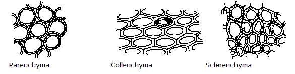
The division and differentiation of the cells of meristematic tissues given rise to permanent tissues. Cell division is the formation of two or more daughter cells from one mother cell. Simple permanant tissue consist of similar permanent cells that perform the same function or a similar set of functions. Parenchyma, collencyma and sclerenchyma are three types of simple permanent tissues. Complex permanent tissue are a group of different types of cells that perform a common function. Xylem and phloem are two types of complex permanent tissue.
* Parenchyma
This tissue is composed of large, thin-walled cells which are generally oval or spherical. The cells are not packed closely, i.e., there are intercellular space. These living cells with a nucleus and a vacuole are found in the soft parts of the plant. They store food, fill up spaces between other tissue and provide temporary support to the plant. When they contain chloroplasts, as in leaves, they help manufacture food.
* Collenchyma
This tissue is composed of cells that are elogated and thickened with cellulose at the corners. There is no intercellular space. Collenchyma provides mechnical support to plant organs and is found in leaf stalks and below the epidermis of stems. It helps leaves and stems bend without breaking. It provides support, protection and flexibility ot plant organs. It is generally absent in roots.
* Sclerenchyma
This tissue is composes of long, narrow cells whose walls are evenly thickened with lignin. Lignin is a chemical that acts like cement, sticking fibres and hardening them. Sclerenchyma cells are dead. They are packed together closely, and provide strength and flexibility to plant parts. They are present in stems, veins of leaves, the hard covering of seeds and nuts, and the husk of coconut. Fibre-yielding plants like jute and flax contain this tissue in abundance.
* Xylem, or wood, as it is often called, is a complex tissue. The cells are thick-walled, tubular and often dead. This tissue has four types of cells– tracheids, vessels, xylem parenchyma and xylem fibres. Of these only tracheids and vessels transport sap.
* Phloem
Phloem too is a complex tissue made up of four types of cells, or elements–sieve tubes, companion cells, phloem fibres and phloem parenchyma. It is not necessary for the phloem to contain all four types of cells. Phloem to contain all four types of cells. Phloem cells are living cells (except phloem fibres) which help transport food from leaves to the storage organs and growing regions of the plant.
’* Animal Tissues
While doing the activities in this chapter, you have come across two types of animal tissue, the cheek cells are a type of epithelial tissue, while blood is a kind of connective tissue. There are two other types of animal tissue–muscular and nervous.
* Epithelial Tissue:
This tissue covers the surface of the body and lines the internal organs. Its main function is protection. The cells that form the different types of epithelial tissue differ in shape. Some are thin and flat, some cubelike, while others are columnar.
* Connective Tissue:
Blood, bones cartilages, tendons (which connect muscles with bones) and ligaments (which tie bones together) are diferent types of connective tissue. Adipose tissue, or what is generally known as fat, is also a kind of connective tissue. Though different in structure, connective tissues have one thing in common-the cells are suspended or embedded in a matrix. In blood, the matrix is liquid and is called the plasma.
’* Muscular Tissue:
Muscular tissue is also of different types. However, the different types of muscular tissue (or muscles) have the same basic functoin. They contract and relax to make different parts of the body move. The muscles in our arms, legs thighs, back and so on help us move. The muscles in the heart help it pump blood. The muscles in the alimentary canal help the passage of food. The muscles in the blood vessels help them dilate and get constricted.
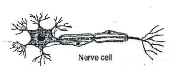
’* Nervous Tissue:
Nerve cells make up nervous tissue. A nerve cell has a long tail and short branches coming out of it.The tail too has branches. These help to carry message from one cell to the other. The brain and spinal cord are make up of nervous tissue.
Differences between prokaryotic & Eukaryotic cells
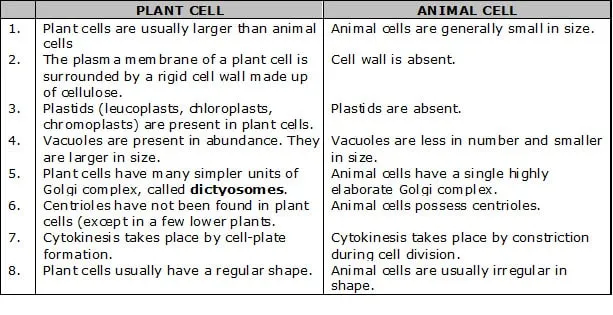
Differences between Plant cell & animal cell
 `
`
NCERT QUESTIONS WITH SOLUTIONS
Q.1 Make a sketch of the human nerve cell. What funtion do nerve cells perform?
Ans. Functions of human nerve cell:
(i) Nerve cells receive message from different parts of body.
(ii) They further transfer these messages to brain and accordingly brain send commands for functioning of different organs of body.
Q.2 Write short notes on the following:
(i) Cytoplasm (ii) Nucleus of a cell
Ans. (i) Cytoplasm: Cytoplasm is a jelly like substance which is present between the cell membrane and the nucleus. Various other organelles of cells are present in the cytoplasm. Cytoplasm is made up of chemical substances like carbohydrates, proteins and water. These chemical substances are present in cells of all types and sizes. Cytoplasm contains many important tiny substances called
Organelles.
(ii) Nucleus of a cell: Nulceus is the master of the cell. It commands all the functioning of the cell. It is generally located in the center of the cell and is spherical in shape. A membrane called nuclear membrane separates it from cytoplasm. It contains the genetic material DNA and RNA in it. This porous membrane allows the transfer of material in the nucleus and cytoplasm. Nucleus contains a dense body called Nucleolus which actually contains Chromosomes, the genetic material.
Q.3 Which part of the cell contains organelles?
Ans. Cytoplasm.
Q.4 State a difference between eukaryotes and prokaryotes.
Ans. Prokaryotes do not have a well designed nuclear membrane while, eukaryotes have a well designed nuclear membrane.
Q.5 Where are the chromosomes found in cell? State their functions?
Ans. Chromosomes are found in the nucleus of a cell. Their function is to carry characteristic features of parent cells to the daughter cell means, from parent to offspring.
Q.6 Cells are the basic structural units of living organism. Explain.
Ans. In Biology, the basic unit of which all living thins are composed is knows as cell. The cell is the smallest structural unit of living matter that is capable of functioning independently. A single cell can be a complete organism in itself, as in bacteria and protozoans. A unicellular organism also captures and digests food, respires, excretes, grows, and reproduces. Similar functions in multi-cellular organisms are carried out by groups of specialized cell which are organized into tissues and organs such as, the higher plants and animals. Hence, ‘cell’ is known as the basic structural and functional unit of life.
Q.7 Explain why chloroplast are found only in plant cells.
Ans. Chloroplasts are found only in plant cells because they are required for photosynthesis.
EXERCISE - I
Q.1 Name the four types of animal tissue
Q.2 What is the importance of ribosomes?
Q.3 What is the function of mitochondria?
Q.4 Name the following:
(a) structural and function unit of life
(b) powerhouse of the cell
Q.5 Who discovered the cell and when ?
Q.6 Name two multicellular organisms.
Q.7 What are pseudopodia ?
Q.8 Mention three different shapes of cells in human body.
Q.9 Which part of the cell gives the shape to a cell.
Q.10 What are chromosomes ?
Q.11 Write the name of unit of inheritance in livings.
Q.12 Name the part of cell which help in control of the activities.
Q.13 Write the name of pigment found in chloroplasts.
Q.14 Why is the plasma membrane called selectively permeable
Q.15 Why lysosomer are called suicidal bags?
Q.16 How many types of organisms on the basis of number of cells ?
Q.17 What are tissues ?
Q.18 Why are mitochondria called the power house of the cell?
Q.19 What are the basic differences between plant cells and animal cells?
Q.20 Name the structural unit of an organism.
Q.21 Write the functions of cell wall.
Q.22 Explain the position and functions of a nucleus in a cell.
Q.23 How is rough ER different from smooth ER? What functions do they perform in a cell?
EXERCISE - II
Q.1 Centriole is associated with –
(A) DNA synthesis
(B) Reproduction
(C) Spindle formation
(D) Respiration
Q.2 The cell organelle associated with cell secretion is
(A) Plastids (B) Mitochondria (C) Golgi apparatus (D) Nucleolus
Q.3 Which of the following is an inclusion?
(A) Mitochondrion (B) Lysosome
(C) Golgi complex (D) Starch grain
Q.4 Which of the following would not be considered part of a cell's cytoplsm?
(A) Ribosome (B) Nucleus
(C) Mitochondrion (D) Microtubule
Q.5 Which of the following is called the brain of the cell?
(A) Nucleus (B) Mitochondria (C) Ribosomes (D) Plasma membrane
Q.6 Which one is not a part of nucleus?
(A) Chromatin (B) Nucleolus
(C) Centrosome (D) Nucleoplasm
Q.7 The common feature amongst nucleus, chloroplast and mitochondrion is –
(A) DNA (B) Lamellae
(C) Cristae (D) All of these
Q.8 Nucleus is separated from surrounding cytoplasm by a nuclear envelope which is –
(A) Single and porous
(B) Double and porous
(C) Single and nonporous
(D) Double and nonporous
Q.9 Nucleoplasm is continuous with cytoplasm through –
(A) Centriole
(B) Golgi apparatus
(C) Nuclear pores
(D) Endoplasmic reticulum
Q.10 Nucleolus was discovered by
(A) Fontana (B) Schleiden
(C) Altmann (D) Robert Brown
Q.11 The function of the nucleolus in the cell is
(A) Secretory
(B) Synthesis of DNA
(C) Synthesis of RNA and ribosomes (D) None of these
Q.12 Which of the following phenomena is commonly referred as 'cell drinking'?
(A) Exocytosis
(B) Pinocytosis
(C) Endocytosis
(D) Phagocytosis
Q.13 The cell organelle taking part in photorespiration is:
(A) Glyoxysome
(B) Dictyosome
(C) Peroxisome
(D) Endoplasmic reticulum
Q.14 Endoplasmic reticulum sometime contains –
(A) Ribosomes (B) Lysosomes (C) Golgi bodies (D) None of these
Q.15 Ribosomes are composed of –
(A) 1 subunit (B) 5 subunits (C) 2 subunits (D) 4 subunits
Q.16 Double membrane is absent in –
(A) Mitochondrion (B) Chloroplast (C) Nucleus (D) Lysosome
Q.17 Animal cell is limited by–
(A) Plasma membrane
(B) Shell membrane
(C) Cell wall
(D) Basement membrane
Q.18 The radiant energy of sunlight is converted to chemical energy and stored as –
(A) AMP (B) ADP
(C) ATP (D) APP
Q.19 Root hair absorbs water from soil through –
(A) Osmosis (B) Active transport (C) Diffusion (D) Endocytosis
Q.20 The barrier between the protoplasm and outer environment in a plant cell is –
(A) Cell membrane (B) Nuclear membrane (C) Cell wall (D) Tonoplast
Q.21 An animal cell differs from a plant cell in respect of –
(A) ER (B) Cell wall
(C) Ribosomes (D) Cell membrane.
Q.22 If the nucleus is a cell's "control centre" and chloroplasts its "solar collectors". Which of the following might be called the cell's combination "food processor" and "garbage disposer"?
(A) Lysosome (B) Ribosome
(C) Golgi apparatus (D) Nucleolus
Q.23 The longest cell in human body is –
(A) Neuron (B) Muscle fibre (C) Epithelial cell (D) Bone cell
Q.24 Identify human cells which lack nucleus–
(A) WBC (B) RBC
(C) Platelets (D) Nerve cells
Q.25 The energy currency of a cell is –
(A) ADP (B) AMP
(C) ATP (D) CTP
Q.26 Which organelle releases oxygen?
(A) Ribosome (B) Golgi apparatus (C) Mitochondria (D) Chloroplast.
Q.27 The term "protoplasm" to the living substance present inside the cell, was given by
(A) Robert Hooke
(B) Robert Brown
(C) J.E. Purkinje
(D) W.Flemming
Q.28 Ribosomes are the centre for –
(A) Respiration
(B) Photosynthesis
(C) Protein synthesis
(D) Fat synthesis.
Q.29 Lysosomes are the reservoirs of
(A) Fat
(B) RNA
(C) Secretory glycoproteins
(D) Hydrolytic enzymes.
Q.30 The membrane surrounding the vacuole of a plant cell is called
(A) Tonoplast
(B) Plasma membrane
(C) Nuclear membrane
(D) Cell wall
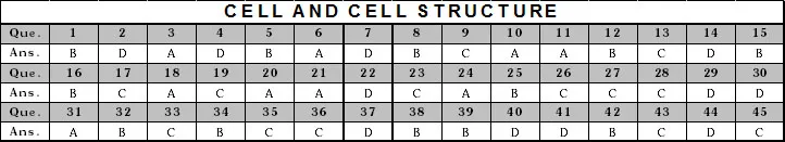
ANSWERS KEYS
1. C 2. C 3. D 4. B
5. A 6. C 7. A 8. B
9. C 10. A 11. C 12. B
13. C 14. A 15. C 16. D
17. A 18. C 19. A 20. C
21. B 22. A 23. A 24. B
25. C 26. D 27. C 28. C
29. D 30. A
EXERCISE - III
CELL AND CELL STRUCTURE
1. The main function of a plasma membrane is to
(A) prevent water from entering or leaving (B) control what goes into and out of the cell
(C) act as a sieve, allowing only lipids to pass (D) move the cell from place to place
2. A cell has the following molecules and structures: enzymes, DNA, ribosomes, plasma membrane, and mitochondria. It could be a cell from
(A) a bacterium (B) an animal, but not a plant
(C) a plant, but not an animal (D) a plant or an animal
3. Cell theory was propounded by
(A) Schleiden and Schwann (B) Watson and Crick
(C) Mendel and Morgan (D) Wallace and Darwin
4. The figures of cork cells as seen by Robert Hooke were published in his book
(A) Origin of Species (B) Plant Kingdom (C) Genra Plantarum (D) Micrographia
5. Who defined protoplasm as a physical basis of life?
(A) Dujardin (B) Huxley (C) Watson (D) Schwann
6. The term 'protoplasm' was given by
(A) Purkinje (B) Dujardin (C) Brown (D) Fischer
7. Which of the following is a prokaryotic cell?
(A) Spirogyra (B) Amoeba (C) Rhizopus (D) Escherichia coli
8. Who first coined the word "cell"?
(A) Aristotle (B) Hooke (C) Schwann (D) Leeuwenhoek
9. Which two organelles are thought to have originated from free-living prokaryotic cells?
(A) Mitochondria and ribosomes (B) Chloroplast and nucleus
(C) Chloroplast and mitochondria (D) Lysosomes and mitochondria
10. Cell wall is
(A) permeable (B) semipermeable (C) differentially permeable (D) impermeable
11. Membranes occur in
(A) cytoplasm, chloroplasts and mitochondria (B) cytoplasm, nuclei and starch grains
(C) chromosomes, chloroplasts and starch grains (D) nuclei, chromosomes and mitochondria
12. Ribosome was discovered by
(A) Porter (B) Palade (C) Muller (D) Ochoa
13. All are membrane bound cell organelles except
(A) mitochondria (B) spherosomes (C) ribosomes (D) lysosomes
14. The smallest organelles in a cell are
(A) lysosomes (B) spherosomes (C) peroxysomes (D) ribosomes
15. Proteins that are to be used outside the cell are synthesised
(A) in the mitochondria (B) on the rough endoplasmic reticulum
(C) on the smooth endoplasmic reticulum (D) on free ribosomes
16. If cells are broken up and sedimented by centrifugation, the new structure formed in one of the fractions is
(A) lysosome (B) microsome (C) ribosome (D) centrosome
17. Golgi apparatus is absent in
(A) liver cells (B) higher plants (C) blue green algae (D) yeast
18. Which of the following is not the correct pairing of structure with function?
(A) Golgi complex: breakdown of complex molecules (B) Mitochondrion: production of ATP
(C) Endoplasmic reticulum: synthesis of proteins (D) Chloroplast: photosynthesis
19. Dictyosomes are
(A) place of flagellar organelle (B) respiratory particle
(C) golgi bodies (D) class of ribosomes
20. Which of the following structure is the functional unit in a Golgi complex?
(A) Cisternae (B) Thylakoid (C) Archoplasm (D) Cristae
21. Major function of Golgi body is
(A) secretion (B) regulation of cell temperature
(C) active transport (D) transportation
22. Cellulose and hemicellulose are the constituents of cell wall are synthesised by
(A) microbodies (B) smooth endoplasmic reticulum
(C) lysosomes (D) Golgi apparatus
23. The enzymes for Kreb's cycle in mitochondria are located
(A) in perimitochondrial space (B) on Inner membrane
(C) in mitochondrial matrix (D) on the outer membrane
24. The presence of DNA in mitochondria and chloroplast supports the hypothesis that
(A) Mitochondria and chloroplast both originated as independent free-living organisms
(B) Glycolysis occurs in both mitochondria and chloroplast
(C) Chloroplast and mitochondria undergo meiosis and mitosis independent of nucleus
(D) ATP is produced in chloroplast as well as mitochondria
25. Green pigments capable of capturing the energy of sunlight are located within the
(A) endoplasmic reticulum (B) chloroplasts (C) cell wall (D) ribosomes
26. Chloroplasts are considered as self-replicating units as they contain
(A) DNA (B) RNA (C) both DNA and RNA (D) neither DNA or RNA
27. When green tomatoes turn red then
(A) new chloroplast are made
(B) chromoplasts are changed into chloroplasts
(C) chloroplasts are disintegrated and get converted into chromoplasts
(D) none of these
28. Which of the following pairs is mismatched?
(A) Nucleus; ribosomal RNA (B) Nucleus; DNA replication
(C) Lysosome; protein synthesis (D) Cytoskeleton; microtubules
29. The filaments that specialise in moving organelles are
(A) micro filaments (B) intermediate filaments (C) actin (D) microtubules
30. Spindle fibres are made up of
(A) lipids (B) cellulose (C) pectin (D) proteins
31. Function of centriole is
(A) formation of spindle fibres (B) formation of nucleolus
(C) initiation of cell division (D) formation of cell plate
32. Chromosomes having equal arms are known as
(A) telocentric chromosome (B) metacentric chromosome
(C) concentric chromosome (D) acrocentric chromosome
33. Electron microscope has revealed the presence of
(A) chloroplasts (B) leucoplasts (C) ribosomes (D) chromosomes
34. The term mitosis was given by
(A) Farmer (B) Flemming (C) Boveri (D) Moore
35. The role of mitosis is not merely to divide a cell into two daughter cells but to ensure genetic continuity from one cell generation to another cell generation. The mechanism ensuring genetic continuity is
(A) formation of cells with new chromosomes (B) formation of two daughter cells
(C) formation of two cells with identical DNA
(D) halving the chromosome number between the two new cells
36. Colchicine interferes in
(A) chromosome condensation (B) DNA replication
(C) organisation and orientation of spindle elements (D) none of these
37. The main significance of mitosis is that
(A) it causes genetic variation (B) it causes transformation of DNA to RNA
(C) it reduces the number of chromosomes (D) it ensures genetic homogeneity of cells
38. The chronological sequence of stages in prophase of meiosis is
(A) Leptotene, Pachytene, Zygotene, Diakinesis, Diplotene
(B) Leptotene, Zygotene, Pachytene, Diplotene, Diakinesis
(C) Zygotene; Leptotene, Pachytene, Diakinesis, Diplotene
(D) Diplotene, Diakinesis, Pachytene, Zygotene, Leptotene
39. Mitochondria are more in cells where
(A) There is least cellular activity (B) There is maximum cellular activity.
(C) There is an average activity (D) They form tissues.
40. The cell theory states all the following except that
(A) Cells are units of structure in plants and animals (B) Cells are units of function
(C) Cells arise from pre-existing living cells (D) Cells always mutate.
41. The main difference between prokaryotic and eukaryotic cell is that the prokaryotic cell does not have
(A) A plasma membrane (B) A cell wall
(C) A genetic system (D) A well defined nucleus with a membrane.
42. The sum total of all processes carried out in a cell is called
(A) Symbiosis (B) Metabolism (C) Anabolism (D) Catabolism
43. The endoplasmic reticulum in a cell probably functions in the
(A) Protein synthesis (B) Photosynthesis
(C) Transport of materials (D) Secretory activities of the cell
44. The two acids present in the nucleus are
(A) Ribonucleic acid and citric acid (B) Nitric acid and malic acid
(C) Deoxyribonucleic acid and fatty acid (D) Ribonucleic acid and deoxyribonucleic acid.
45. Most cells lacking chloroplasts do not have
(A) Ribosomes (B) Mitochondria (C) Cell wall (D) Cell membrane.
1. A tissue is a :
(A) group of separate organs that are coordinated in their activities
(B) group of similar cells that function together in a specialised activity
(C) layer of cells surrounding an organ
(D) sheet of cells, one layer thick
2. Endothelium of the inner surface of blood vessels in vertebrates is formed
(A) simple squamous epithelium
(B) columnar epithelium
(C) cuboidal epithelium
(D) ciliated cells
3. Cells of squamous epithelium are
(A) columnar (B) tall with elongated nuclei
(C) flat plate-like (D) cube like
4. The protein deposited in the dead superficial cells that make the skin epithelium impervious to water is
(A) keratin (B) elastin (C) collagen (D) mucus
5. Horns of most mammals are composed of
(A) bones (B) cartilage (C) keratin (D) chitin
6. Mammary glands are modified
(A) sebaceous gland (B) sweat gland
(C) oil gland (D) lymph gland
7. Which of the following tissue has matrix, which is the source of its structural and functional performances?
(A) connective tissue (B) muscular tissue
(C) nervous tissue (D) epithelial tissue
8. Wrinkling in old age is due to
(A) collagen (B) myosin (C) keratin (D) actin
9. Dermis of mammalian skin is mainly composed of
(A) muscular tissue (B) epithelial tissue (C) connective tissue (D) all of the above
10. Fat is abundant in
(A) liver cells (B) alveolar tissue (C) lymph glands (D) adipose tissue
11. Tendon connects
(A) cartilage with muscles (B) bone with muscles
(C) ligament with muscles (D) bone with bone
12. Cartilage is produced by
(A) osteoblasts (B) epithelium (C) fibroblasts (D) chondroblasts
13. Bone forming cells are
(A) osteoblasts (B) osteoclasts (C) chondroblasts (D) chondroclasts
14. Bone marrow is important for
(A) breakdown of WBC (B) production of RBC
(C) breakdown of RBC (D) production of WBC
15. Haversian system is typically found in bones of
(A) fishes (B) aves (C) reptiles (D) mammals
16. Blood is
(A) acidic (B) alkaline (C) variable (D) neutral
17. Mammalian erythrocytes are
(A) circular (B) biconcave
(C) non-nucleated (D) all the above
18. Life span of RBC is
(A) 50 days (B) 75 days
(C) 120 days (D) 100 days
19. Red cell count is carried out by
(A) haemocytometer (B) haemoglobinometer
(C) sphygmomanometer (D) electrocardiogram
20. Striated muscles are found in
(A) gall bladder (B) wall of bronchi
(C) leg muscles (D) lungs
21. Smooth muscles occur in
(A) vein (B) artery (C) uterus (D) all the above
22. Cardiac muscle is made of branched fibres that are
(A) nonstriated and under voluntary control
(B) striated and not under voluntary control
(C) nonstriated and not under voluntary control
(D) striated and under voluntary control
23. Junction of two neurons is called
(A) synapse (B) junction (C) synapsis (D) synapticula
24. Most of the human neurons are
(A) multipolar (B) bipolar
(C) unipolar (D) pseudo-unipolar
Plant Tissue
1. Prosenchyma is a type of :-
(A) Chlorenchyma (B) Collenchyma (C) Parenchyma (D) Sclerenchyma
2. Tracheary elements are :-
(A) Suberised (B) Full of dense cytosol
(C) Elongated with lignified walls (D) Small and numerous
3. The function of vessels/xylem is to :-
(A) Translocate water and minerals
(B) Translocate organic food
(C) Translocate water alone
(D) Translocate water, minerals and provide mechanical strength
4. Resin ducts of Pinus are an example of :-
(A) Lysigenous cavity
(B) Schizogenous cavity (C) Vacuolated cells
(D) Intercellular spaces
5. Enucleate thin-walled cells with perforated septa are :-
(A) Prosenchyma
(B) Sieve cells (C) Sieve tube elements (D) Collenchyma
6. Vascular bundle having xylem and phloem on the same radius is :-
(A) Concentric (B) Radial (C) Open (D) Collateral
7. The growth in plants is :-
(A) Cuased by each and every cell of the body (B) Limited to certain regions
(C) Caused by non-dividing regions (D) Uniform
8. Aerenchyma occurs in :-
(A) Mesophytes (B) Xerophytes (C) Hydrophytes (D) Sciophytes
9. Angiosperms contain :-
(A) No sieve tubes (B) No vessels (C) Tracheids only (D) Vessels
10. One of the following tissue is responsible for cell division in plants :-
(A) Meristematic tissue (B) Xylem (C) Phloem (D) Sclerenchyma
11. Which group possesses vessels in its xylem :-
(A) Pteridophytes (B) Angiosperms (C) Gymnosperms (D) Both B and C
12. Collenchyma occurs in the stem and petioles of
(A) Xerophytes (B) Monocots (C) Dicot herbs (D) Hydrophytes
13. Which is correct about transport or conduction of substances :-
(A) Organic food moves upwardly through xylem
(B) Organic food moves up through phloem
(C) Inorganic food moves upwardly and downwardly through xylem
(D) Organic food moves upwardly and downwardly through phloem
14. A bicollateral vascular bundle is characterised by :-
(A) Phloem being sandwitched between xylem (B) transverse splitting of vascular bundle
(C) Longitudinal splitting of vascular bundle (D) Xylem being sandwitched between phloem
15. Sieve tube differs from a vessel in :-
(A) Absence of nucleus (B) Absence of cytoplasm (C) Absence of lignification (D) Absence of plasmodesmata
16. Lignin occurs in the cell walls of :-
(A) Phloem (B) Cork
(C) Woody tissue/Xylem cells (D) Parenchyma/Epidermal cells
17. Collenchyma occurs in :-
(A) Herbaceous climbers (B) Woody climbers
(C) Climbing stems (D) Water plants
18. Aerenchyma assists the plants in :-
(A) Attachment (B) Mechanical strength (C) Floating on water (D) Exchange of gases
19. When parenchyma contains chlorophyll and performs photosynthesis, it is called :-
(A) Chlorenchyma (B) Collenchyma (C) Prosenchyma (D) Aerenchyma
20. Which one is not a plant fibre :-
(A) Coir (B) Flax (C) Hemp (D) Silk
21. The only plant cells without nucleus among the following is :-
(A) cambium cells (B) root hair (C) companion cells (D) xylem vessels
22. Trachea, tracheids, wood fibres, and parenchymatous tissues are found in :-
(A) xylem (B) cambium (C) cortex (D) phloem
23. Collenchyma differs from parenchyma in having
(A) cellulose walls (B) vacuoles
(C) pectin deposits at corners (D) living protoplasm
24. A tissue whose living cells form the mechanical tissue of activity growing organs and whose cell walls show cellulosic unlignified thickenings often at the corners of its cells is called :-
(A) sclerenchyma (B) collenchyma (C) Chlorenchyma (D) parenchyma
25. Sieve tubes are better suited for translocation because these :-
(A) are broader than long (B) possess bordered pits
(C) possess no end walls (D) possess a broader lumen and perforated cross walls
26. Companion cells are usually seen associated with
(A) fibres (B) parenchyma (C) xylem vessels (D) sieve tubes
27. Grass stem elongates by the activity of :-
(A) secondary meristem (B) intercalary meristem (C) apical meristem
(D) primary meristem
28. Guard cells are present in :-
(A) Stomata (B) Cortex (C) Cork (D) Vascular bundle
29. Vascular bundles in the stem of Cucurbita are :-
(A) radial (B) collateral (C) bicollateral (D) concentric
30. Secondary growth occurs through :-
(A) Formation and division of meristematic cells (B) Vascular region
(C) Cortical region (D) Both 2 and 3
31. In which of the following, growth is subapical?
(A) root (B) shoot (C) pedicle (D) petiole
32. Polyarch condition is seen in :-
(A) monocot root (B) dicot root (C) dicot stem (D) monocot stem
33. Vascular bundle in dicot roots is :-
(A) conjoint (B) radial (C) collateral (D) bicollateral
34. Well-developed pith is found in :-
(A) monocot stem and dicot root (B) monocot root and dicot stem
(C) dicot root and dicot stem (D) monocot root and monocot stem
35. In root, xylem is referred as :-
(A) polyarch (B) exarch (C) endarch (D) mesarch
36. In dicot root :-
(A) vascular bundles are arranged in ring and have cambium
(B) xylem and phloem radially arranged
(C) xylem is always endarch
(D) vascular bundles are scattered
37. A tracheid differs from a vessel in having :-
(A) scalariform thickenings (B) discontinuous lumen, which are separated by end wall
(C) lack of bordered pits (D) thick walls without contents
38. Vessels and companion cells are characteristic of xylem and phloem of :-
(A) pteridophytes (B) gymnosperms (C) angiosperms (D) bryophytes
39. Collenchymatous hypodermis is present in :-
(A) dicot stem (B) monocot as well as in dicot stems
(C) monocot stem (D) monocot root
40. Phloem parenchyma is absent in :-
(A) dicot leaf (B) monocot stem (C) dicot stem (D) dicot root
41. Cork cells are :-
(A) photosynthetic (B) elongated and participate in movement
(C) meristematic (D) dead
42. In which of the following phloem occurs in two patches?
(A) bicollateral (B) collateral (C) amphicribral (D) amphivasal
43. Husk of coconut is made of :-
(A) Parenchyma (B) Sclerenchymatous tissue
(C) Collenchyma (D) Prosenchyma
44. Dendrochronology is the study of :-
(A) height of a tree
(B) age of a tree by counting the number of annual rings in the main stem
(C) diameter of a tree
(D) age of tree by counting the number of leaves in the main stem
45. The youngest layer of secondary phloem in woody dicot stem is located :-
(A) Just outside the vascular cambium
(B) Just on the inner side of cambium
(C) Between periderm and primary cortex
(D) Just outside pith
46. The wall of cork cells is mostly impregnated with
(A) Cutin (B) Suberin (C) Lignin (D) Hemicellulose
47. Mesophyll tissue is well differentiated into spongy tissue and palisade tissue in :-
(A) dicot leaves (B) xerophytic stem (C) hydrophytic stem (D) monocot leaves
48. What is the other name for the cork tissue?
(A) phellogen (B) phelloderm (C) phellem (D) periderm
49. The manufactured food in green plants moves from the leaves to other parts through :-
(A) pith (B) xylem (C) cortex (D) phloem
50. Sieve tubes have :-
(A) simple oblique wall (B) perforated and longitudinal plates
(C) perforated and oblique septa (D) apical and oblique plates