
The cell and its structures are studied under a branch of biology called cytology.
Definition :- The structural & functional unit of living beings is called cell. 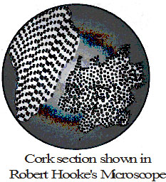
1. Robert Hooke (1665) :– An English man and first curator of
Royal society of London.
Observed a thin transverse section of bark of a tree under self designed microscope.
He noticed honey - comb like compartments.
He coined the term cell .
He wrote a book - Micrographia.
He actually observed dead cells.
2. Antony Van Leeuwenhoek (1674) was first to observe
living cells like bacteria [from tartar of teeth]
erythrocytes [fish], sperms and protozoans [eg. Vorticella]
3. N. Grew (1682) :– Proposed cell concept which states that cell is unit of structure of organisms.
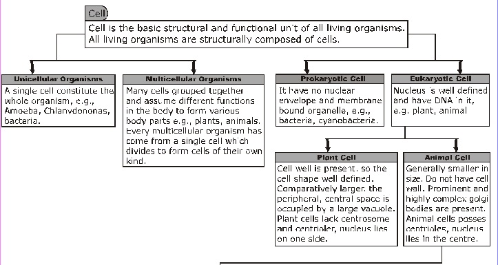
4. Cell is called structural & functional unit of life because –
(i) All the living organisms are composed of one or more cells.
(ii) All the cells have similar basic structure.
(iii) Similar cell organelles of different cells perform similar functions.
5. Knoll and Ruska (1932) of Germany designed the electron microscope which was employed to study the ultrastructrue (fine structure) of cell and various cell organelles in 1940s.
It is instrument which is used to study those objects that cannot be seen with the naked eye or with the help of a hand lens. A microscope has more than one lens. The 1st compound microscope was built by F. Janssen and Zacharias Janssen (1590).
² Structure of Microscope: The microscope used in schools is called compound microscope, a compound microscope has following parts:
1 Base: It is the basal, metallic, horse-shoe shaped structure. It bears the whole weight of microscope.
2 Handle: It is the curved part to hold the microscope. It is also called as arm.
3 Stage: It is a strong metallic, rectangular, horizontal plate fixed to the handle.
4.Stage Clips: Two clips are attached to stage used for holding the slide in position.
5.Condenser: Below the stage is present a condenser for concentrating the light rays.
6. Body tube: It is wide, hollow tube attached to the upper part of the arm. To this tube lenses are attached.
7.Adjustment Screw:
(a) Coarse adjustment: It is bigger sized screw used to move the body tube up and down.
(b) Fine adjustment: It is a smaller sized screw for fine focussing.
8 Reflecting Mirror: It is meant for reflecting the light rays, so that light passes through the object which is to be seen.

Two biologists, "Schleiden and Schwann' gave the "Cell theory" which was later on expanded by "Rudolf Virchow". Cell theory states that-
(i) All plants and animals are composed of cells.
(ii) Cells is the basic unit of life.
(iii) All cells arise from pre-existing cells.
Ciruses are the exceptions of cell theory.
(A) Size of cell – Normal size in human 20 µm to 30 µm in diametre.
(i) Largest cell – In animals – Ostrich egg [15 cm is diametre]
In plants – Acetabularia [6-10 cm]
(ii) Longest cell – In animals – Nerve cell [upto 1mt]
In plants – Hemp fibre.
(iii) Smallest cell – PPLO – Pleuro Pneumonia Like Organism [Mycoplasma – 0.1 to 0.5 µm.]
(B) Shape of cell – Shape of cell mainly depends upon the specific function it performs.
(i) Elongated – Nerve cell (ii) Discoidal/saucer – RBC
(iii) Spindal – Muscle cell (iv) Spherical – Eggs.
(v) Branched – Pigment cell of the skin. (vi) Slipper shaped – Paramecium
(vii) Cuboidal – Germ cells of gonads. (viii) Polygonal – Liver cells.
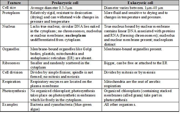
(A) On the basis of type of organization, cells are of two types:
(i) Prokaryotic cells: these are primitive and incomplete cells. they have less developed nucleus without nuclear membrane and nucleolus e.g. Bacteria.
(ii) Eukaryotic cells: these are well developed cells. They have advanced nucleus with nuclear membrane.
(B) On the basis differentiation:
(i) Undifferentiated: These are unspecialized cells which by mitotic divisions give rise to new cells for the formation and maintenance of tissues.
(ii) Differentiated: These are specialized cells formed from the unspecialized cells by change in structure and function during develoment and growth of an organism.
(iii) Dedifferentiated: These are specialized cells reverted to a more generalized (embryonic), actively dividing state. Dedifferentiation often occurs for regeneration.
There is an occurrence of division of labour within a cell as they all got certain specific components called "Cell organelles" each of them performs a specific function.
The three basic components of all the cells are
(i) PM (Plasma Membrane) (ii) Nucleus (iii) Cytoplasm
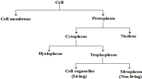
· Mitochondria · Vacuole
· Endoplasmic Reticulum · Starch granules
· Golgibody · Store food materials
· Plastid · Wastes
· Lysosome
· Ribosome
Each cell (prokaryotic as well as eukaryotic) is surrounded by a covering called plasma membrane or plasmalemma or cell membrane. Most cell organelles in eukaryotic cells (e.g., Mitochondria, Plastids, Golgi apparatus, Lysosomes, Endoplasmic reticulum, Peroxisomes, Vacuoles etc). are enclosed by subcellular unit membranes. These membranes, thus, compartmentalise the cell.
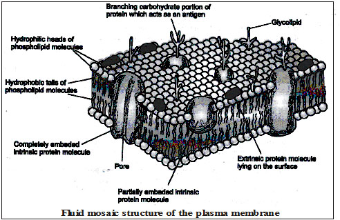
Molecular Structure of Plasma membrane.
Plasma membrane is a living, ultra-thin, elastic, selectively permeable membrane. Chemically, it is composed of phospholipids, proteins, oligosaccharides and cholesterol.
Trilamilar or 3-layered structure :- J.D. Robertoson noted trilamilar or 3-layered structure for all membranes he studied. Based on his findings, he proposed the 'unit membrane hypothesis' in 1959.
Fluid Mosaic Model :- In 1972, S.J. Singer and G. Nicolson proposed fluid mosaic model to explain the structure and functions of plasma membrane. According to this model, the plasma membrane is made up of a phospholipid bilayer and two types of protein molecules 'floating about' in the fluid phospholipid bilayer. The two types of proteins are (i) Intrinsic proteins which are embeded in the phospholipid matrix incompletely or completely, and (ii) Extrinsic proteins which occur superficially either on the outer surface or on the inner surface of the phospholipid layer. In other words, the membrane is a viscous fluid with phospholipids and protein molecules arranged as a mosaic.
Oligosaccharide molecules are present on the exposed surface of the plasma membrane. They are associated with proteins as well as lipid molecules forming glycoproteins and glycolipids respectively. Cholesterol molecules are inserted between the phospholipid molecules of plasma membrane of animal cells to stabilize the membrane.
Presence of lipids and proteins provides flexibility to the plasma membrane. Proteins present in the membrane serve as :-
(i) Enzymes catalysing chemical reactions within the membrane.
(ii) Transport proteins (permeases) for movement of water soluble ions.
(iii) Pumps for active transport of materials and
(iv) Receptor proteins (e.g., glycoproteins on the cell surface) to recognize and bind specific molecules such as hormones.
Fluid mosaic model is also described as "a number of protein icebergs floating in the sea of lipids'.
Types of membranes :-
(i) Impermeable membrane :- If the membrane does not allow passage of substances (solvent and solute) through it.
(ii) Permeable membrane :- If the membrane allows free passage of solute and solvent through it.
(iii) Semipermeable membrane :- If the membrane allows passage to solvents but prevents the passing of solutes.
(iv) Selectively permeable membrane :- If the membrane allows the passage of solvent and few selected
solutes.
Advantage of Semipermeability membrane :- Semipermeability ensures that
1. The useful molecules enter the cell,
2. The metabolic intermediates remain within the cell and
3. The secretions and wastes leave the cell.
Thus, semipermeability of cell membranes enables the cell to maintain homeostasis, i.e., a constant internal environment inspite of the changes outside it.
The substances generally drawn in the cell include :
(i) Raw materials for metabolism, viz. food stuffs, water, salts and oxygen; and
(ii) Regulatory substances, e.g., vitamins and hormones.
The substances generally turned out of the cells include :
(i) The products of metabolism, namely, nitrogenous wastes and carbon dioxide; and
(ii) Secretions.
Following mechanisms are involved in the entry or exit of various materials across p.m.
(A).Physical processes. (B) Biological processes.
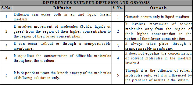
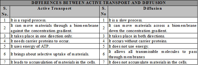
A.Physical Processes :- These processes are slow and do not expend energy. These occur down the concentration gradient and do not use carrier proteins. Physical processes include. (i) Diffusion, (ii) Osmosis.
B.Biological processes :- These processes are rapid and often use energy in the form of ATP. These can occur down as well as against the concentration gradient and often use carrier proteins. Biological processes include:-
1. Mediated transport
(i) Facilitated transport / diffusion (ii) Active transport
2. Endocytosis (Pinocytosis and Phagocytosis)
3. Exocytosis.
1 Diffusion :- The process by which a substance uniformly spreads into another substance by random movement of its particles from a region of higher concentration to a region of its lower concentration due to their kinetic energy is called diffusion.
It is faster in gaseous phase than in liquid phase or solid phase.
Significance of diffusion :-
(i) Diffusion helps in the distribution of various substances throughout the cytoplasm of the cell without much delay.
(ii) It helps in the exchange of respiratory gases (oxygen and carbon dioxide) between the body cells and their environment.
(iii) Various materials such as gases, liquids and solids dissolve in the medium, i.e., air or liquid by diffusion.
(iv) Loss of water in vapours form from the aerial parts of the plants (transpiration) occurs through diffusion.
(v) Flowers of plants spread aroma through diffusion. It attracts insects and other animals for pollination.
Each cell (prokaryotic as well as eukaryotic) is surrounded by a covering called plasma membrane or plasmalemma or cell membrane. Most cell organelles in eukaryotic cells (e.g., Mitochondria, Plastids, Golgi apparatus, Lysosomes, Endoplasmic reticulum, Peroxisomes, Vacuoles etc). are enclosed by subcellular unit membranes. These membranes, thus, compartmentalise the cell.
Plasma membrane is a living, ultra-thin, elastic, selectively permeable membrane. Chemically, it is composed of phospholipids, proteins, oligosaccharides and cholesterol.
Trilamilar or 3-layered structure :- J.D. Robertoson noted trilamilar or 3-layered structure for all membranes he studied. Based on his findings, he proposed the 'unit membrane hypothesis' in 1959.
Fluid Mosaic Model :- In 1972, S.J. Singer and G. Nicolson proposed fluid mosaic model to explain the structure and functions of plasma membrane. According to this model, the plasma membrane is made up of a phospholipid bilayer and two types of protein molecules 'floating about' in the fluid phospholipid bilayer. The two types of proteins are (i) Intrinsic proteins which are embeded in the phospholipid matrix incompletely or completely, and (ii) Extrinsic proteins which occur superficially either on the outer surface or on the inner surface of the phospholipid layer. In other words, the membrane is a viscous fluid with phospholipids and protein molecules arranged as a mosaic.
Oligosaccharide molecules are present on the exposed surface of the plasma membrane. They are associated with proteins as well as lipid molecules forming glycoproteins and glycolipids respectively. Cholesterol molecules are inserted between the phospholipid molecules of plasma membrane of animal cells to stabilize the membrane.
Presence of lipids and proteins provides flexibility to the plasma membrane. Proteins present in the membrane serve as :-
(i) Enzymes catalysing chemical reactions within the membrane.
(ii) Transport proteins (permeases) for movement of water soluble ions.
(iii) Pumps for active transport of materials and
(iv) Receptor proteins (e.g., glycoproteins on the cell surface) to recognize and bind specific molecules such as hormones.
Fluid mosaic model is also described as "a number of protein icebergs floating in the sea of lipids'.
Osmosis :-
The diffusion of water or solvent through a semipermeable membrane from a solution of lower concentration of solutes to a solution of higher concentration of solutes to which the membrane is relatively impermeable, is called osmosis.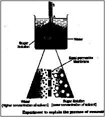
Osmosis is of two types :
1. Endomosis
2. Exomosis
Endosmosis : It is the entry of water molecules into the cells through semipermeable plasma membrane when surrounded by hypotonic solution.
Exosmosis : It is the exit of water molecules from the cells through semipermeable plasma membrane when surrounded by hypertonic solution.
Experiment : Demonstration of osmosis in the laboratory.
Requirements : Funnel fitted with a semipermeable membrane, beaker, sugar solution, water.
Procedure : Take sugar solution in a funnel fitted with a semipermeable membrane (fish bladder or egg membrane) upto mark 'A' and place it in an inverted position in a beaker filled with clean water as shown in figure. After some time, observe the level
of sugar solution in the funnel. Result :- You would find that the sugar solution has risen from level
'A' to a new level 'B'.
Explanation and conclusion : Sugar solution in the funnel and water in the beaker are separated by a semipermeable membrane. The fitted membrane is permeable to small water molecules but is relatively impermeable to large sugar molecules dissolved in water.
Due to difference in the concentration of solute on the two sides of semipermeable membrane, water molecules have moved from the solution having lower concentration of solutes (e.g., water in this experiment) to the solution having higher concentration of solutes [e.g. sugar solution] due to osmosis has risen to new level 'B'.
Types of solutions :
1. Isotonic solution
2. Hypotonic solution, and
3. Hypertonic solution.
1.Isotonic solution :- Isotonic solution is one in which the concentration of water and solutes is the same as in the cytoplasm of the red blood cells. 0.9% salt solution and 5% glucose solution are isotonic for red blood cells.
2. Hypotonic solution :-Hypotonic solution is one in which the concentration of solutes is less and concentration of water is more as compared to inside the red blood cells. 0.66% salt solution and 0.2% glucose solution are hypotonic for red blood cells.
3. Hypertonic solution :-Hypertonic solution is one in which the concentration of solutes is more and the concentration of water is less as compared to in the cytoplasm of the red blood cell. 1.25% salt solution and 10% glucose solution are hypertonic for red blood cells.
Other examples of osmosis :-
1. Fresh water unicellular organisms (e.g., Amoeba, Paramecium) continuously gain water in their bodies due to osmosis. These organisms have mechanisms (e.g., contractile vacuoles) to throw out excess of water from their bodies.
2. Most plant cells have the tendency to gain water due to osmosis.
3. Absorption of water by the plant roots from the soil through root hairs is also an example of osmosis.
4. Certain plant movements (e.g., seismonastic movements in 'touch-me-not' plant) occur due to loss or gain of water.
5. Stomata are present in the leaves. They open and close at different times of the day due to osmotic movements of water.
6. In plants, cells, tissues and soft organs (leaves, young shoots, flowers) maintain turgidity or stretched form due to osmotic absorption of water.

Mediated transport :
Type of transport of materials across the 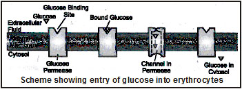
plasma membrane with the help of carrier
proteins is called mediated transport.
Types of mediated transport
Mediated transport is of following two types :
(i) Facilitated transport
(ii) Active transport
(i) Facilitated transport :- In this case, transport proteins (e.g. permeases) assist molecules to diffuse through the membrane down the concentration gradient, i.e., from the region of higher concentration to the region of lower concentration across the membrane. It is, therefore, also termed as facilitated diffusion. No cellular energy is used in such transport. A carrier protein combines with a specific substance (e.g., glucose) to be transported and moves it down the concentration gradient from one side of membrane to another through a channel formed by it.
In liver and red blood cells, facilitated transport moves glucose across the cell membrane by specific carrier protein molecule in both directions, depending upon whether glucose concentration is higher inside or outside the membrane.
(ii) Active transport :- In this case, carrier proteins move substances against the concentration gradient, i.e., from lower concentration to higher concentration. This "uphill" transport involves work and always requires energy provided by ATP (adenosine triphosphate).
Mechanism of active transport of materials is described below :
(i) The carrier protein has a binding site for ATP in addition to the binding site for the substrate. As the ATP molecule binds to the carrier protein, it is hydrolyzed to ADP.
(ii) The energy so set free brings the substrate binding site of the carrier protein to the surface of the membrane. The substrate present in the medium joins the carrier protein at substrate binding site to form carrier-substrate complex.
(iii) The substrate bond carrier protein undergoes conformational change and carries the substrate through a channel in it to the cytoplasmic side of the membrane.
(iv) Now, the form of binding site changes and the substrate is released. The carrier protein regains its original form and is ready to transport another molecule of substrate.
There are many active transport systems in the cell. Among these, sodium-potassium exchange pump is prominent. It maintains sodium and potassium gradients between cells and the surrounding extracellular fluid.
Importance of active transport :- The Na+ – K+ exchange pump plays following roles :
(i) It helps in maintaining a positive charge on the outside of the membrane and negative charge on the inside (resting potential),
(ii) It helps in nerve impulse conduction,
(iii) It helps in muscle contraction,
(iv) It helps in urine formation in kidney tubules,
(v) It helps in salt excretion in marine birds, and
(vi) It helps in controlling water contents of the cell.
Animal cells can also actively take in and turn out materials in masses much larger than in the hither to described processes by utilizing energy. Such materials include macromolecules, lipid droplets and solid particles. Items of this size cannot cross the phospholipid bilayer by diffusion or with the help of transport proteins. Special processes are involved in the transport of such large quantities of materials.
These include endocytosis (phagocytosis) and exocytosis.
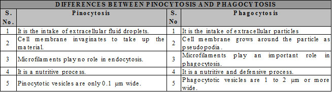
The term endocytosis refers to invagination of a small region of plasma membrane, and ultimately forming an intracellular membrane-bound vesicle. Endocytosis is not shown by plant cells because of their rigid cell wall and internal turgor pressure. Depending upon the intake of fluid droplet or solid particles, endocytosis is of two types :
(i) Pinocytosis (ii) Phagocytosis
(i) Pinocytosis :- The non-specific intake of a tiny droplet of extracellular fluid by a cell through the cell membrane which cannot otherwise pass through it. It is also, therefore, termed as cell drinking. It was first observed in Amoeba. In this process, a small region of plasma membrane invaginates and the fluid droplet passes into the pocket so formed. This pocket is called caveola. The pocket deepens and finally nips off as a fluid-filled vacuole called pinosome or pinocytotic vesicle.
(ii) Phagocytosis :- Phagocytosis is the intake of solid particles by a cell through cell membrane. It is also called cell eating. Phagocytosis is the major feeding method in many unicellular organisms (e.g., Amoeba) and simple metazoa (e.g., sponges).
An area of the plasma membrane, coated initially with actin-myosin, comes in contact with the food particle(s). The contact induces the cell membrane to put out tiny protoplasmic processes, the pseudopodia, around the food particle(s). The pseudopodia meet on the other side of the food particle(s) and fuse. In this way, an internal vacuole, called phagosome, containing food particle(s) in a droplet of water is acquired.
Exocytosis :-
Exocytosis is the process that involves fusion of membrane of the exocytotic vesicle with the plasma membrane to extrude its contents to the surrounding medium.
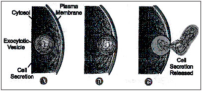
This process is also called cellular vomiting or ephagy and the vesicles that turn out the materials are termed exocytotic vesicles.
Exocytosis process is responsile for :
(i) removal of undigested food left in the food vacuoles in the cells.
(ii) secretion of substances such as hormones, enzymes, and
(iii) replacement of internalized membrane by the fusion of exocytotic vesicles with the cell membrane.
1. It gives a definite shape to the cell.
2. It provides protection to the internal contents of the cell.
3. It regulates entry and exit of substances in and out of the cell.
4. It can internalize solid and liquid materials by infolding or extending around them. This is a process of active intake of materials.
5. In animal cells, it is involved in adhesion, recognition and in the formation of vesicles, cilia, flagella, microvilli, etc.
Plasma membrane acts as a mechanical barrier to protoplasm so after rupturing or breakdown of
plasma membrane, the protoplasmic contents will be dispersed in the surrounding medium.
Discovered by Robert Hooke
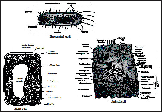
(i) The outermost covering of the plant cell is called cell wall.
(ii) It is absent in animal cell.
(iii) It is rigid, thick, porous and non-living structure. It become impermeable due to deposition of cell wall materials.
Middle lamella : Common layer between two plant cells is called middle lamella. It consists Ca & Mg pectates (Plant cement). Fruits becomes soft and juicy due to dissolve of middle lamella.
(i) 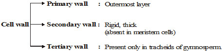
(ii) Cellulose is a main constituent of cell wall but addition to cellulose – Hemicellulose, cutin, pectin, Lignin, Suberin are also presents in cell wall
(iii) Cellulose microfibrils and macrofibrils arranged in layers to form skeleton of cell wall. In between these layers other substances like pectin, hemicellulose may be present. These form matrix of cell wall.
(iv) Network of cellulose fibre forms skeleton of cell wall.
35-100 cellulose chain = 1 micelle.
20 micelle = 1 Microfibril
250 micro fibril = 1 macrofibril in cell wall.
(v) Composition:– (i) Cellulose + Hemicellulose-in plants
(ii) Chitin – in fungi
(iii) Peptidoglycan – in bacteria and mycoplasma.
Functions of cell wall :–
1. It determines the shape of the plant cell.
2. It prevents desiccation of cell. [desiccation means drying up of cells]
3. It protects the plasma membrane and internal structures of the cell.
4. It helps in the transport of various substances in and out of the cell.
5. It does not allow too much of water to come in. In this way it prevents the cytoplasm from becoming too dilute.
1. Cytoplasm was discovered by Kolliker in 1862.
2. It is the site of both biosynthetic and catabolic pathways.
3. It can be divided into two parts:
(i) Cytosol: Aqueous soluble part contains various fibrous proteins forming cytoskeleton.
(ii) Cytoplasmic Inclusion: In the cell cytoplasm, there are present numerous living and
non-living structures, collectively called cytoplasmic inclusions.
(iii) Cytoplasmic Inclusion: In the cell cytoplasm, there are present numerous living and
non-living structures, collectively called cytoplsmic inclusions.
(a) The living cytoplasmic inclusions are called cell organelles or protoplasmic inclusions or organolds and
(b) the non-living structures are called Deutoplasmic or ergastic bodies.
(i) Participates in intracellular distribution of nutrients, metabolites and enzymes.
(ii) Helps in exchange of materials between cell organelle.
(iii) acts as a site of chemical reactions like glycolysis (step of respiration), synthesis of fatty acids.
Introduction :
(i) The nucleus is the most important component of the cell and controls all functional activities of the cell.
Historical Account :
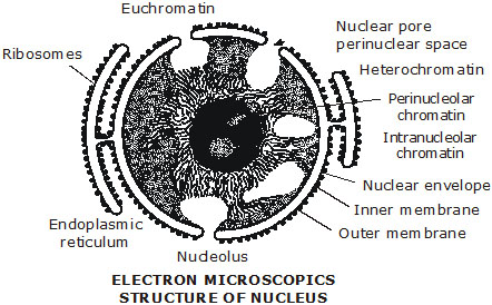
(i) Robert Brown (1831) discovered a dense, spherical body in the cells of an ‘orchid’ and named it as ‘Nucleus’.
(a) Nuclear envelope : Nucleus is surrounded by two membranes, that separates nucleoplasm from cytoplasm. The nuclear membrane has minute pores. These are called nucleo-pores.
(b) Nucleoplasm : The part of protoplasm which is enclosed by nuclear membrane is called nucleoplasm. It contains chromatin threads and nucleolus.
(c) Nucleolus : Discovered by Fontaina. Usually one nucleolus is present in each nucleus but sometimes more than one nucleoli are present. It is a store house of RNA.
(d) Chromation threads : A darkly stained network of long and fine threads called chromatin threads. Chromatin threads are intermingled with one another forming a network called chromation reticulum. Whenever the cell is about to divide the chromatin material gets organized into chromosomes.
Functions of Nucleus :
(i) The nucleus control all metabolic activities of the cell.
(ii) It regulates the cell cycle.
(iii) It brings about growth of the cell by directing the synthesis of structural proteins.
(iv) It takes part in the formation of ribosomes.
(v) It contains genetic information and is concerned with the transmission of hereditary traits from one generation to another.
Do you know?
Chromatin threads are made up of –
(i) DNA (ii) Protein [Histone protein]
Gene:– The segment of DNA and act as unit of heredity
ATP:– Adenosine triphosphate. It is also known as energy currency. It provides energy to perform bio-synthesis & mechanical work.
Homologous chromosomes:– All chromosomes are found in pair and the chromosomes of a pair are called homologous chromosomes.
Non-homologous chromosomes:– Chromosomes of different pair.
The nucleus of prokaryotes is also known nucleoid.
Nucleus is also called director of cell as it controls most of the cellular activities.
Nucleus is absent in sieve tubes of vascular plants & mature RBC's of mammals. Mammalian
RBC also lacks Golgibodies, mitochondria, ER, lysosomes.
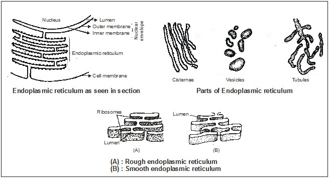
(i) In the cytoplasm some closed or open, branched cavities are present which are bounded by membranes to form a network of membranous system called Endoplasmic Reticulum.
(i) K.R.Porter (1948) reported this net-like system under electron microscope.
(i) A system of membranes attached to the nucleus and present in the cytoplasm is called E.R.
(ii) The Endoplasmic Reticulum (ER) is divided into two parts
² It is the netowork of membranes present in the cytoplasm.
² It was discovered by Porter, Claude and Fullan.
² These are present in all cells except prokaryotes and mammalian erythrocytes.
² They are made up of three components:
(A) Cisternate: These are long, flattened, parallely arranged, unbranched tubules.
These form successive layers of nucleus.
These are found in cells which are active in protein synthesis and are 40 - 50 mm in diameter.
(B) Vesicles: These are rounded or spherical, They are found in synthetically active cells.
(C) Tubules: These are small, smooth walled and have tubular spaces. These are found in non secretory as well as steroid synthesizing cells.
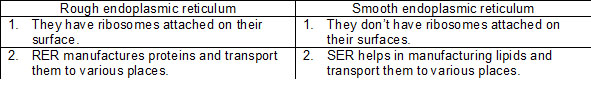
(a) Rough Endoplasmic Reticulum (RER)
(b) Smooth Endoplasmic Reticulum (SER)
(i) RER possesses rough wall because ribosomes remain attached on the surface. RER is present in cells which are involved in protein synthesis.
(ii) SER mainly present in cells which are involved in lipoproteins and glycogen synthesis. It perfoms detoxification.
q Functions of Endoplasmic Reticulum :
(i) It forms supporting skeleton framework of the cell.
(ii) Certains enzymes present in smooth E.R. synthesis fats (lipids), steroids and cholesterol.
(iii) Rough E.R. is concerned with protein synthesis.
(iv) Smooth E.R. is involved in the process of detoxification.
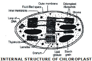
It is a double membranous discoidal structure, found only in plant cells.
Chloroplast was discovered by A.V. Leeuwenhoek and named by Schimper.
Besides being discoidal or rhombic in plant cells they occur in variable shapes like in algae they can be 'U' shaped, spiral, coiled, ribbon shaped etc.
In each thylakoid Quantasomes are present which are called as Photosynthetic units.
Each quantasome possesses 230 chlorophyll molecules.
Each chloroplast consists of two parts.
(i) Grana: It constitutes the lamellar system. These are found layered on top of each other, these stacks are called as Grana.
Each granum of the chloroplast is formed by superimposed closed compartments called Thylakoids.
Functions: Grana are the sites of light reaction of photosynthesis as they contain phtosynthetic pigment chlorophyll.
(ii) Stroma: It is a granular transparent substance also called as matrix.
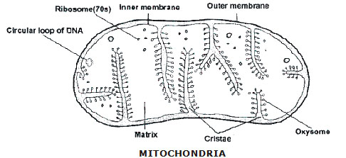
1 It was first seen by Kolliker in insect cells and named by Benda.
2 It is a rod shaped structure found in cytoplasm of all eukaryotic cell except mammalian RBC's.
3 These are also absent in prokaryotes.
4 Maximum mitochondria are found in metabolically active cells.
5 It is also called as "Power House of the Cell" or the "Storage Battery".
6 It is double membranous structure where outer membrane has specific proteins while inner me:nbrane is folded inside to form chambers called Cnstae."Cristae" are the infoldings of inner mitochondrial membrane that possess enzymes for respiratory cycles like Kreb Cycle. ATP synthesizing units are called Oxysomes or F0 – F1 Particles.
7 Space between inner and outer mitochondrial membranes is called as perimitochondrial space. The fluid present in mitochondria is called as matrix.
(a) Functions:
(i) Its main function is to produce and store the energy in the form of ATP.
(ii) It is the site of Kreb's cycle of respiration, as it contains enzymes for Kreb cycle.
(iii) Oxysome contains enzymes for ATP production.
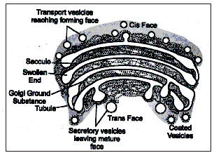
Discovered by Camillo Golgi (1898) in nerve cells of owl.
Other names:–
(i) Lipochondrion, (ii) Idiosome,
(iii) Baker's body, - In fungus (iv) Dalton complex
(v) Dictyosomes – In plants
Position:– It is located near the nucleus.
· The cytoplasm surrounding Golgi body have fewer or no other organelles. It is called Golgi ground substance or zone of exclusion.
· Golgi bodies are pleomorphic structures, becaue component of golgi body are differ in structure & shape in different cells.
Structure:– It is formed of four types of contents.
(i) Cisternae – These are long flattened and unbranched saccules. 4 to 8 saccules are arranged in a stack.
(ii) Tubules – These are branched and irregular tube like structures associated with cisternae.
(iii) Vacuoles – Large spherical structures associated to tubules.
(iv) Vesicles – Spherial structures arise by budding from tubules. Vesicles are filled with secretory materials.
Golgibody is single membrane bound cell organelle.
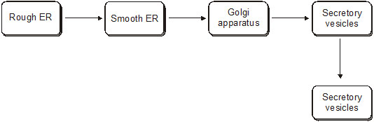
Function:–
(i) It involved in cell-secretion and acts as storage, modification and condensation or packaging membrane.
(ii) It forms the Acrosome of sperm [Acrosome :- A bag like structure filled with lytic enzymes which dissolve egg membrane at the time of fertilization]
(iii) It forms the lysosomes and secretory vesicles.
(iv) It is the site for formation of glycolipids and glycoproteins.
(v) Synthesis of cell wall material (Polysaccharide synthesis)
(vi) Cell plate formation (phragmoplast) during cell formation.
(vii) Vitelline membrane of egg is secreted by Golgi body.
First observed and the term coined by Christian De Duve (1955)
· Lysosomes are spherical bag like structures [0.1 – 0.8 µm] which is covered by single unit membrane.With the exception of mammalian RBC they are reported from all cells. Lysosomes are filled about 50 different types of digestive enzymes termed as acid hydrolases.
· Lysosomes are highly polymorphic cell organelle. Because, during functioning, lysosomes have different morphological and physiological states.
· Primary lysosomes or storage granules – These lysosomes store enzyme Acid Hydrolases in their inactive form. These are newly formed lysosome.
· Digestive vacuoles or Heterophagosomes – These lysosome forms by the fusion of primary lysosomes and phagosomes. These are also called secondary lysosomes.
· Residual bodies – Lysosomes containing undigested material are called residual bodies. These may be eliminated by exocytosis. These are also called as Telolysosomes. (Tertiary lysosomes)
· Autophagic lysosomes or cytolysosomes or autophagosomes – Lysosomes which digest cell organelles are known as Autophagosomes.
(i) Heterophagy :– It involve in digestion of foreign materials received in cell.
(ii) Autophagy :– Digestion of old or dead cell organelles.
(iii) Cellular digestion (Autolysis) :– Sometimes all lysosomes of a cell burst to dissolve the cell completely.
That's why lysosomes also known as suicidal bags.
Chemically a ribosome is made of proteins and RNA. 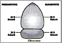
² First reported by Claude and named by G.P alade.
² They are small granular structures visible only under electro microscope.
² They are the only organelles which are present in all types of cells.
² They help in protein synthesis and are known as 'protein factories'.
² Each ribosomes consists of two unequal subunits, larger dome shaped and small ovoid.
The size of ribosome is determined by sedimentation coefficient in the centrifuge.
The cytoplasmic ribosomes of eukaryotes are 80S and in prokaryotes and cell organelles like mitochondria and chloroplast it is 70S type. The two sub units of 80S ribosomes are 60S and 40S while 70S type ribosomes have 50S and 30S subunits.
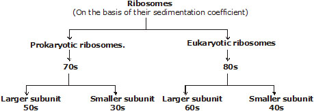
· Magnesium ion [Mg++] is essential for binding of both the sub units of ribosome.
Functions :–
Site of protein synthesis, so these are also called protein factories.
Peroxisomes/Uricosomes.
· Discovered by Rhodin & Tolbert.
· Peroxisome term was first used by De Duve.
It contains per-oxide forming enzymes.
Functions :–
(i) In animals peroxisomes are concerned with b-oxidation of fatty acids & peroxide metabolism.
(ii) In plants peroxisomes are concerned with b-oxidation of fatty acids, peroxide metabolism and
photorespiration.
· Scattered Golgibodies in the cytoplasm of plant cells are also called Dictyosomes.
· Lysosome found in four forms that's why it is also called polymorphic cell organelle.
· Chloroplasts are centres of photosynthesis to prepare the organic food so are called kitchens of the cells.
· Vacuoles of animal cells arise from Golgi-complex.
· Tonoplast:– Plasma membrane that covers the vacuole is called tonoplast.
Vacuoles are of three types :-
1. Food vacuole – The vacuole which contain food material.
2. Sap vacuole – The vacuole which is filled by liquid material [sap]
3. Contratile vacuole – The vacuole that concern with osmoregulation e.g. Amoeba
Functions :–
(i) Storage of food, water and other substences.
(ii) They help in the elimination of excess water from the cell (osmoregulation), and maintains internal pressure of the cell
Centrosome :– Discovered by Benden. Boveri named it as centrosome.
· Centrosome is generally found in animal cells. Only few type of a plant cells show its presence.
· It is situated near the nucleus of the cell and shaped like star.
· Each centrosome has two centrioles. The two centrioles are placed perpendicular to each other.
· Cytoplasm which surrounds centrioles called as "Centrosphere". Centrioles and centrosphere collectively called centrosome or microcentrum or diplosome.
(i) In animal cells centrioles play important role in initiation of cell division by arranging spindle fibres between two poles of cell.
(ii) The location of centrioles during cell division decides the plane of division.
(iii) It form the basal granule of cilia and flagella in micro-organisms, zoo-spores & motile gametes.
(iv) Form tail of sperm.
(i) In many eukaryotic as well as prokaryotic cells of both plants and animals a cytoskeleton has been reported in recent years.
(ii) The elements of this cytoskeleton are proteins.
(iii) The cytoskeleton consists of following two elements within a cell.
(a) Microtubules
(b) Microfilaments
(iv)Cilia and flagella of eukaryotic cells are microscopic, contractile & filamentous process of cytoplasm.
(v) Cilia is shorter than flagella and are numerous.
(A) Microtubules :
Introduction :
(i) These are cylindrical structures formed by the polymerization of two-part subunits of globular protein tubulin into helical stacks.
Historical Account :
The term ‘microtubule’ was coined by Slautterback in 1963.
Ultrastructure :
(i) Microtubles radiate from each end of the cell. Which helps in the movement of chromosomes.
(ii) These are found in many plant and animal cells.
Function :
(i) Microtubules help in the structure and movement of cillia and flagella.
(ii) It also play a role in cell division.
(B) Microfilaments :
Ultrastructure :
(i) These are long and helically intertwined polymers. Microfilaments are made up of protein actin.
Function :
(i) These filaments help in cell movement and in formation of cell furrow and cell plate.
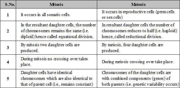
(i) Cell multiplication is needed for the growth, development and repair of the body. Cell multiplies by dividing itself again and again this process called cell division.
(ii) Cell divisions are two types
(a) Mitosis (b) Meiosis
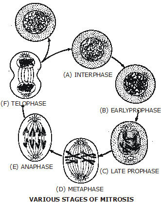
Stages of Mitosis :
Interphase, prophase, metaphase, anaphase and telophase are roughly the five stages or phases of mitosis.
(a) Interphase :
(i) The period between one cell division and the next is called interphase in which the cell is said to be in the resting stage.
(ii) Interphase, however, includes three phases, i.e. G1-phase, S-phase and G2-phase. G1-phase is a resting phase or pre-DNA synthesis phase.
(iii) During S-phase, DNA synthesis takes place. G2-phase is again a resting phase and it may be described as a post-DNA synthesis phase.
(iv)The main mitosis division takes place during M-phase which involves prophase, metaphase, anaphase and telophase.
(b) Prophase :
(i) Prophase is actually the first and the longest phase in the mitosis cell division.
(ii) Chromosomes become visible in the nucleus as short, thick and helically-coiled threads.
(iii) Each chromosome splits into two chromatids joined at the centromere.
(iv)Nuclear membrane dissolves away.
(v) Nucleolus also dissolves away and finally disappears.
(c) Metaphase :
(i) It is the second stage in the mitotic cell division.
(ii) Nuclear membrane and nucleolus disintegrate and they are lost completely.
(iii) Spindle tubules start appearing, and these tubules get attached to chromosomes at the centromeres.
(iv) Chromosomes move actively, become shorter and thicker and arrange themselves in the centre or on the equator of the spindle.
(v) Separation of the two chromatids from each chromosomes also begins at the end of metaphase.
(d) Anaphase :
(i) It is the third stage of mitosis.
(ii) Chromatids separate from each other at centromeres.
(iii) Separated sister chromatids, each with a centromere, are called daughter chromosomes. They move to the ends of opposite poles of the spindle.
(iv)Daughter chromosomeres appear in V, U or J-shaped during their movement towards the poles.
(v) During the late anaphase stage, the cell starts constricting in the middle region.
(e) Telophase :
(i) Telophase is the last stage of mitotic cell division.
(ii) Chromatids or daughter chromosomes are now at the end of the spindle.
(iii) Nuclear membranes and nucleoli reform around each group of chromosomes and thus two new nuclei are reorganized at each pole.
(iv)Chromosomes begin to lose their compact structure.
(v) Spindle Xapparatus disappears gradually.
Division of nucleus is called karyokinesis and, the process of the division of cytoplasm is called cytokinesis.
(i) In animal cells, a circular constriction appears at the equator, the constriction deepens and eventually divides the cell into two.
(ii) In plant, there is no constriction. A cell plate or new cell wall forms across the cell resulting in the separation of two daughter cells.
Significance of Mitosis :
(i) Mitosis occurs during the growth and development of multicellular plants and animals.
(ii) Mitosis ensures that the two daughter cells inherit the same number of chromosomes.
(iii) It helps the cell in maintaining proper size.
(iv)In unicellular organisms mitosis helps in asexual reproduction during which two or more individuals arise from the mother cell.
(v) If mitosis becomes uncontrolled it may cause tumour or cancerous growth.
(i) Meiosis is also called reduction division because the chromosomes in this division are reduced from the diploid to the haploid number.
(ii) Meiosis occurs in all organisms which reproduce sexually.
(iii) Meiosis produces haploid sex cells from diploid cells.
(iv) Meiosis involves two cell division, viz., meiosis I and meiosis II.
(v) In meiosis I, the replicated homologous chromosomes pair with each other on the spindle, cross over and then separate to either end of the spindle.
(vi) On the other hand, in meiosis II, the chromatids of each chromosome move towards the centromere, and these chromatids separate at each end of the second spindle.
(vii)As a result of this process, a diploid cell divides to form four haploid cells.
First Meiosis Division :
First meiosis division is actually the reduction division. It consists of prophase I, metaphase I, anaphase I and telophase I.
(a) Prophase I :
(i) Prophase I is the longest phase of meiosis and includes five sub-phases.
(i) Leptotene :
(i) This is the first stage in the first meiosis prophase.
(ii) In this stage, the chromosomes appear as separate thin and fine thread-like structures.
(ii) Zygotene :
(i) Homologous chromosomes come together, or arrange themselves side by side in pairs to form bivalents.
(ii) This pairing of homologous chromosomes during zygotene in the first meiosis prophase is called synapsis.
(iii) Pachytene :
(i) The bivalents or chromosomes become shorter and thicker.
(ii) They replicate or split into chromatids but remain linked at the centromeres.
(iii) Each bivalent thus now consists of four chromatids.
(iv) Crossing over between non-sister chromatids of homologous pair takes place.
(iv) Diplotene :
(i) The centromeres of paired chromosomes or bivalents move away from each other and crossing over can also be seen.
(ii) The points in a bivalent where the two chromosomes appear to be joined and crossed over are called chiasmata.
(iii) Chiasmata formation and crossing over are the distinguishing features of diplotene.
(v) Diakinesis :
(i) This is the last stage of first meiosis prophase.
(ii) The chromosomes become shortest and thickest.
(iii)Terminalisation of chiasmata.
(iv)Nuclear membrane starts disintegrating. Nucleolus also disintegrates. Diakinesis followed by metaphase I.
(b) Metaphase I :
(i) Nuclear membrane disappears completely at the beginning of metaphase I.
(ii) Pairs of homologous chromosomes are lined up at the centre.
(iii) Spindle apparatus starts appearing. Few spindle fibres get attached with the centromeres of chromosomes.
(iv)Metaphase I change into anaphase I.
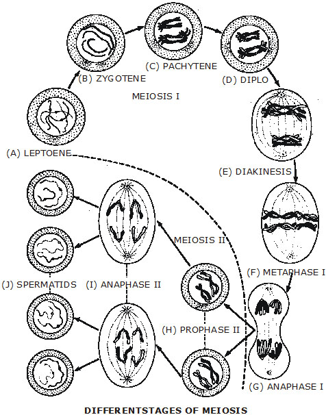
(c) Anaphase I :
(i) Partners of homologous chromosomes separate completely and move to opposites poles of spindle during anaphase I, which in turn changes into telophase I.
(d) Telophase I :
(i) The separated partners of homologous chromosomes collect at the poles of the spindle and nuclear membranes form around them. Two daughter haploid nuclei are thus formed. The chromosomes lengthen as they uncoil. Nucleoli start reappearing.
Second Meiosis Division :
Like mitosis, the second meiosis divisions also consists of four phases, i.e. prophase II, metaphase II, anaphase II and telophase II.
Prophase II :
(i) In both the haploid nuclei, each chromosome splits up into two chromatids with a single functional centromere. The nuclear membrane and nucleolus disintegrate partially or completely.
Metaphase II :
(i) The chromatids arrange themselves at metaphase plate or spindle.
Anaphase II :
(i) During anaphase II, the centromere splits. The two chromatids belonging to each chromosomes may now be called chromosomes and pass to the two opposite poles of spindle.
Telophase II :
(i) The haploid set of chromosomes at two different poles of spindle uncoil and form chromatin material. Nuclear membrane forms around each haploid set of chromosomes. Nucleolus also reappears.
Significance of Meiosis :
(i) Meiosis results in the formation of haploid gametes (sperm and ovum)
(ii) The phenomenon of crossing over provides new combinations of chromosomes and, hence new combinations of genes and also of characters in offspring.
(iii)The four chromatids of a homologous pair of chromosomes are passed on to four different daughter cells. This is called the segregation of chromosomes. This causes genetic variations in daughter cells.
(iv)Failure of meiosis leads to the formation of diploid gametes which on fusion form polyploids.
Special Note :
Besides mitosis and meiosis, there is also a third type of division. It is called amitosis. It is a direct division of the nucleus by constriction.
Q.1 Plasma membrane is made up of which two components?
Sol. The two components are lipids and proteins.
Q.2 Cell wall is made up of which components?
Sol. Cell wall is made up of cellulose.
Q.3 Give an example of unicellular organism.
Sol. Amoeba, Bacteria, Paramedium.
Q.4 What is the intracellular source of digestive enzyme?
Sol. Lysosome.
Q.5 What is the function of mitochondria?
Sol. Mitochondria are sites of cellular respiration in which energy, i.e., packets of ATP are formed.
Q.6 Name two structures found in animal cells but not in plant cells.
Sol. Lysosomes and Centrioles.
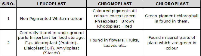
Q.7 Give the name of colourless plastids.
Sol. Leucoplast.
Q.8 What is plasmolysis?
Sol. The shrinkage of protoplasm away from cell wall due to loss of water by osmosis when the cell is kept in hypoertonic medium.
Q.9 What is the function of the cell wall?
Sol. The cell wall lies outside the plasma membrane and is responsible for providing structural strength to the plants.
Q.10 There would be no plant life in chloroplasts did not exist. Justify.
Sol. Chloroplast contains the pigment chlorophyll which is responsible for food preparation by photosynthesis in plants. Hence, if there were no chloroplasts then there would not have been any plant life.
Q.11 Why the Golgi apparatus is called the secretary organelle of the cell?
Sol. This is because it packages material synthesised in the ER and dispatches it to intracellular (plasma membrane and lysosomes) and extracellular (cell surface) targets.
Q.12 Differentiate between smooth and rough endoplasmic reticulum.
Sol. Differences between Smooth and Rough Endoplasmic Reticulum are
Q.13 What is Cytosol and Cytoskeletone?
Sol. Cytosol is the semi-fluid part of the cell cytoplasm which is embedded with organelles. Cytoskeletone is a network of fibres present in the cell which provides a supporting framework for the organelles.
Q.14 What is membrane biogenesis? How plasma membrane is formed during this process?
Sol. The process of plasma membrane formation is called membrane biogenesis.
Q.15 Why are peroxisomes mostly found in kidney and liver cells?
Sol. Peroxisomes contain various oxidative enzymes which detoxify the toxic material.
Since the blood carries various toxic substances to kidney and liver, a large number of peroxisomes are present in them to oxidise the toxid material.
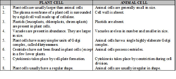
Q.16 What is the difference between plant cell and animal cell?
Sol. 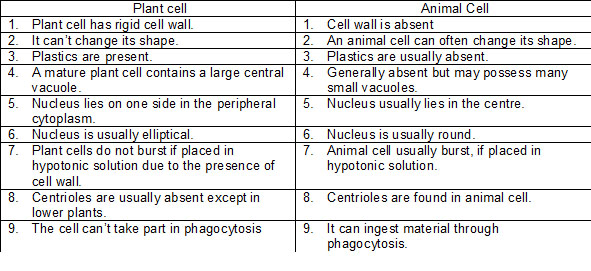
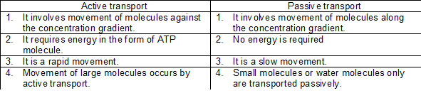
Q.17 What is the active transport? Differentiate between active and passive transport.
Sol. The process in which the molecules are moved uphill against the concentration gradient. Active transport always involves the expenditure of energy because the materials are pumped against the concentration gradient.
Q.1 Who discovered cells and how ?
Q.2 Why the cell is called the structural and functional unit of life ?
Q.3 How substances like carbon dioxide and water move in and out of the cell ?
Q.4 Why is the plasma membrane called a selectively permeable membranes ?
Q.5 Fill in the gaps in the following difference between prokaryotic and eukaryotic cells.
Q.6 Can you mane the two organelles we have studied that contain their own genetic material?
Q.7 If the organisation of a cell is destroyed due to some physical or chemical influence, what will happen ?
Q.8 Why are lysosomes known as suicidal bags ?
Q.9 Where are protein systhesised inside the cell ?
Q.10 Plasmas membrane is made up of which two components ?
Q.11 What is hypotonic solution ?
Q.12 What is hypertonic solution ?
Q.13 What is isotonic solution ?
Q.14 Cell wall is made up of which component ?
Q.15 Give an example of unicellular organism.
Q.16 Give an example of multicelluar organism.
Q.17 What is active transport ?
Q.18 What is the intracellular source of digestive enzyme ?
Q.19 What is endocytosis ?
Q.20 What is the function of mitochondria ?
Q.21 What does ATP stand for ?
Q.22 Which cell organelle is responsible for the release of energy as ATP ?
Q.23 Where are genes located ?
Q.24 Name two structures found in plant cells but not in animal cells.
Q.25 Name two structures found in animal cells but not in plant cells.
Q.26 Give the name of colourless plastids.
Q.27 What is membrane biogenesis ?
Q.28 Which organelle is involved in the formation of lysosomes ?
Q.29 Which organelle is responsible for the storage, modification and packaging of produce in vesiscles ?
Q.30 What is the outermost layer found in animal cells ?
Q.31 What is the outermost layer found in the plant cell ?
Q.32 Which organelle helps in photosynthesis ?
Q.33 Which organelle is the storage sac of solid and liquid materials ?
Q.34 Which organelle serves as a channel for transport of materials between cytoplasm and nucleus ?
Q.35 What is microscope ?
Q.36 Why light microscope is called a compound microscope ?
Q.37 What are cell organelles ?
Q.38 Which organelle digests unwanted organic substances ?
Q.39 Which organelle helps in protein synthesis ?
Q.40 Which organelle is associated with ribosome formations ?
Q.1 Double membrane is absent in –
(A) Mitochondrion (B) Chloroplast (C) Nucleus (D) Lysosome
Q.2 Animal cell is limited by–
(A) Plasma membrane
(B) Shell membrane
(C) Cell wall
(D) Basement membrane
Q.3 The radiant energy of sunlight is converted to chemical energy and stored as –
(A) AMP (B) ADP (C) ATP (D) APP
Q.4 Root hair absorbs water from soil through –
(A) Osmosis (B) Active transport (C) Diffusion (D) Endocytosis
Q.5 The barrier between the protoplasm and outer environment in a plant cell is –
(A) Cell membrane (B) Nuclear membrane (C) Cell wall (D) Tonoplast
Q.6 An animal cell differs from a plant cell in respect of –
(A) ER (B) Cell wall (C) Ribosomes (D) Cell membrane.
Q.7 If the nucleus is a cell's "control centre" and chloroplasts its "solar collectors". Which of the following might be called the cell's combination "food processor" and "garbage disposer"?
(A) Lysosome (B) Ribosome (C) Golgi apparatus (D) Nucleolus
Q.8 The longest cell in human body is –
(A) Neuron (B) Muscle fibre (C) Epithelial cell (D) Bone cell
Q.9 Identify human cells which lack nucleus–
(A) WBC (B) RBC
(C) Platelets (D) Nerve cells
Q.10 The energy currency of a cell is –
(A) ADP (B) AMP
(C) ATP (D) CTP
Q.11 Which organelle releases oxygen?
(A) Ribosome (B) Golgi apparatus (C) Mitochondria (D) Chloroplast.
Q.12 The term "protoplasm" to the living substance present inside the cell, was given by
(A) Robert Hooke (B) Robert Brown (C) J.E. Purkinje (D) W.Flemming
Q.13 Ribosomes are the centre for –
(A) Respiration (B) Photosynthesis (C) Protein synthesis (D) Fat synthesis.
Q.14 Lysosomes are the reservoirs of
(A) Fat
(B) RNA
(C) Secretory glycoproteins
(D) Hydrolytic enzymes.
Q.15 The membrane surrounding the vacuole of a plant cell is called
(A) Tonoplast (B) Plasma membrane (C) Nuclear membrane (D) Cell wall
Q.16 Centriole is associated with –
(A) DNA synthesis (B) Reproduction (C) Spindle formation (D) Respiration
Q.17 The cell organelle associated with cell secretion is
(A) Plastids (B) Mitochondria (C) Golgi apparatus (D) Nucleolus
Q.18 Which of the following is an inclusion?
(A) Mitochondrion (B) Lysosome
(C) Golgi complex (D) Starch grain
Q.19 Which of the following would not be considered part of a cell's cytoplsm?
(A) Ribosome (B) Nucleus
(C) Mitochondrion (D) Microtubule
Q.20 Which of the following is called the brain of the cell?
(A) Nucleus (B) Mitochondria (C) Ribosomes (D) Plasma membrane
Q.21 Which one is not a part of nucleus?
(A) Chromatin (B) Nucleolus
(C) Centrosome (D) Nucleoplasm
Q.22 The common feature amongst nucleus, chloroplast and mitochondrion is –
(A) DNA (B) Lamellae
(C) Cristae (D) All of these
Q.23 Nucleus is separated from surrounding cytoplasm by a nuclear envelope which is –
(A) Single and porous
(B) Double and porous
(C) Single and nonporous
(D) Double and nonporous
Q.24 Nucleoplasm is continuous with cytoplasm through –
(A) Centriole
(B) Golgi apparatus
(C) Nuclear pores
(D) Endoplasmic reticulum
Q.25 Nucleolus was discovered by
(A) Fontana (B) Schleiden
(C) Altmann (D) Robert Brown
Q.26 The function of the nucleolus in the cell is
(A) Secretory
(B) Synthesis of DNA
(C) Synthesis of RNA and ribosomes (D) None of these
Q.27 Which of the following phenomena is commonly referred as 'cell drinking'?
(A) Exocytosis (B) Pinocytosis (C) Endocytosis (D) Phagocytosis
Q.28 The cell organelle taking part in photorespiration is
(A) Glyoxysome
(B) Dictyosome
(C) Peroxisome
(D) Endoplasmic reticulum
Q.29 Endoplasmic reticulum sometime contains –
(A) Ribosomes (B) Lysosomes (C) Golgi bodies (D) None of these
Q.30 Ribosomes are composed of –
(A) 1 subunit (B) 5 subunits (C) 2 subunits (D) 4 subunits
Q.31 In chloroplasts, chlorophyll is present in the–
(A) Stroma (B) thylakoids
(C) Outer membrane (D) Inner membrane
Q.32 The sedimentation coefficient of complete ribosome in bacterial cell is
(A) 70S (B) 80S
(C) 78S (D) 60S
Q.33 Which one of the following is common in plant and animal?
(A) Mitochondria (B) chloroplast (C) Centriole (D) Cell wall
Q.34 Which of the following is a nonliving cell inclusion?
(A) Vacuoles (B) Ribosomes (C) Centrosomes (D) Golgi complex
Q.35 Cell vacuole contains
(A) Water
(B) Metabolic gases
(C) Cytoplasm
(D) Water and dissolved substances
Q.36 A mature plant cell has –
(A) Protoplasm and vacuole
(B) Vacuole and cell wall
(C) Cell wall and protoplasm
(D) Protoplasm, cell wall and vacuole
Q.37 Centriole takes part in –
(A) Cell plate formation
(B) Spindle formation
(C) Nucleolus formation
(D) Start of cell division
Q.38 Which of the following is called 'an organelle within an organelle'? –
(A) Plastid (B) Ribosome
(C) Lysosome (D) Microsome
Q.39 Cell organelle common in Protista and Monera is –
(A) Vacuole (B) Ribosome
(C) Lysosome (D) Chloroplast
Q.40 Which of the following organelles lack membranes?
(A) Ribosome (B) Mitochondria (C) Golgi complex (D) Nucleus
Q.41 Besides cellulose microfibrils, the other two cell wall networks are :-
(A) Protein and hemicellulose
(B) Hemicellulose and protein
(C) Pectin and glycoprotein
(D) Pectin and hemicellulose
Q.42 Middle lamella occurs :-
(A) Inner to primary wall
(B) Inner to secondary wall
(C) Outer to secondary wall
(D) Outer to primary wall
Q.43 Hydrophilic chemical of cell wall is :-
(A) Pectin (B) Suberin
(C) Fat (D) Lignin
Q.44 Structural element of cell wall is :-
(A) Matrix
(B) Microfibrils
(C) Microtubules
(D) Arabinogalactans
Q.45 Different layers of cell wall are :-
(A) Middle lamella and primary wall
(B) Primary wall and secondary wall
(C) Middle lamella, primary wall and secondary wall
(D) Wall layers exclude middle lamella
Q.46 The first wall layer of cell is :-
(A) Tertiary wall, if present
(B) Secondary wall
(C) Primary wall
(D) Middle lamella, if present
Q.47 Plant cells are distinguishable from animal cell in containing :-
(A) Mitochondria (B) Ribosomes
(C) E.R. (D) Cell wall
Q.48 Ripe fruits soften due to :-
(A) Degeneration of cell walls
(B) Partial solubilisation of pectic compounds
(C) Metabolism of tannins
(D) Exosmosis
Q.49 Ribosomes contain large quantities of :-
(A) haemoglobin (B) fatty acid
(C) ribonucleic acid (D) deoxyribonucleic acid
Q.50 Glycocalyx is :-
(A) Glycoproteins and glycolipids
(B) Oligosaccharide part of glycolipids and glycoproteins
(C) Lipid and protein parts of glycolipids (D) Mucopolysaccharides attached to cell wall
Q.51 Which of the following organelles lack membranes ?
(A) Ribosome (B) Lysosome
(C) Golgi body (D) Nucleus
Q.52 Protein synthesis occurs on :-
(A) ribosome (B) nucleus
(C) lysosome (D) centrosome
Q.53 The term protoplasm was coined by :-
(A) Huxley (B) Purkinje
(C) Dujardin (D) Schultze
Q.54 A unit of protoplasm having a nucleus and covered by plasmalemma is called :-
(A) Ectoplast (B) Cell
(C) Cytoplast (D) All the above
Q.55 The term cytoplasm was coined by :-
(A) Sachs (B) Strasburger (C) Hanstein (D) Flemming
Q.56 Which of the following is correct for prokaryotic ribosome :-
(A) it dissociates into 50S and 30S
(B) it dissociates into 40S and 40S
(C) it dissociates into 60S and 20S
(D) it dissociates into 70S and 30S
Q.57 Golgi apparatus takes part in synthesis of :-
(A) Glycolipids (B) Glycoproteins (C) Hormones (D) All the above
Q.58 In a cell DNA is found in :-
(A) nucleus, mitochondria and plastid (B) nucleus, mitochondria and Golgi body
(C) mitochondria, Golgi body and plastid (D) nucleus, Golgi body and plastid
Q.59 Cartilage matrix is digested during its osteogenesis through :-
(A) Intracellular autophagic activity
(B) Extracellular lysosomal activity
(C) Intracellular heterophagic activity (D) Both B and C
Q.60 Which one is lysosomal activity :-
(A) Reabsorption of tadpole tail
(B) Mobilisation of stored substances
(C) Removal of obstructions
(D) All the above
Q.61 When are lysosomes extra-active :-
(A) Seed maturation (B) Seed germination (C) Flowering (D) Fruiting
Q.62 In animal cell, a mitochondrion is :-
(A) Largest organelle
(B) Second largest organelle
(C) Third largest organalle
(D) None of the above.
Q.63 Outer mitochondrial membrane resembles bacterial membrane and outer chloroplast membrane in having :-
(A) Selective permeability
(B) Single ion channels
(C) Porin
(D) All the above
Q.64 Chromoplasts are formed from chloroplasts during :-
(A) Ripening of Tomato
(B) Ripening of Chilli
(C) Development carrot
(D) Both A and B
Q.65 Experiments on Acetabularia by Hammerling proved the role of :-
(A) nucleus in heredity
(B) nucleoplasmic ratio
(C) chromosomes in heredity
(D) cytoplasm in controlling differentiation
Q.66 The plastids with irregular shape are :-
(A) Leucoplasts
(B) Chloroplasts
(C) Chromoplasts
(D) Amyloplasts
Q.67 Peroxisomes and glyoxisomes are :-
(A) Energy transforming organelles
(B) Membrane-less organelles
(C) Macrobodies
(D) Microbodies
Q.68 Structure of nuclear envelope facilitates :-
(A) spindle organization
(B) separation of daughter chromosomes
(C) synapsis of homologous chromosomes (D) nucleocytoplasmic exchange of materials
Q.69 Microfilaments were discovered by :-
(A) Slautterback (B) Paleviz et al (C) Altman (D) Ledbetter and Porter
Q.70 Microfilaments are required for :-
(A) Movement of flagella and cilia
(B) Cell polarity
(C) Sol-gel changes
(D) All the above
Q.71 Cell polarity is determined by :-
(A) Intermediate filaments
(B) Microtubules
(C) Protofilaments
(D) Centrioles
Q.72 Who coined the term 'Nucleolus' ?
(A) Brown (B) Hooke
(C) Fontana (D) Bowman
Q.73 Which of the following phenomena is commonly referred as 'cell drinking' ?
(A) Exocytosis (B) Pinocytosis
(C) Endocytosis (D) Phagocytosis
Q.74 The two centrioles of a pair occur :-
(A) Parallel to each other
(B) At right angles to each other
(C) At an angle other than 90°
(D) End to end
Q.75 Cell organelle having a cartwheel constitution is :-
(A) Centriole and basal body
(B) Microtubule
(C) Microfilament
(D) Basal plate
Q.76 A flagellum beats :-
(A) Independently, undulatory and asymmetrically
(B)Independently, undulatory and symmetrically
(C) Coordinated, pendular and symmetric (D) Coordinated, pendular and asymmetric
Q.77 Food vacuole is formed from :-
(A) Absorbed and digested food
(B) Phagosome + Lysosome
(C) Feeding canals + Lysosome
(D) Feeding canals + Phagosome
Q.78 Chromatin material which remains condensed during interphase is called :-
(A) Heterochromatin (B) Euchromatin (C) Chromonemata (D) Megachromatin
Q.79 Nucleolus was discovered by :-
(A) Robert Brown (B) Leeuwenhoek (C) Robert Hooke (D) Fontana
Q.80 Nucleolus is formed from :-
(A) Nucleus
(B) nuclear sap
(C) Sat chromosome
(D) Giant chromosome
Q.81 Components of nucleus are :-
(A) Karyotheca, nucleolus, chromatin, nucleoplasm and nuclear matrix
(B) Nuclear envelope, nucleolus and chromatin
(C) Nuclear envelope, nucleoplasm and chromatin
(D) All the above
Q.82 Which one of the following pairs is not correctly matched ?
(A) Nucleus - Genetic information
(B) Cell membrane - Permeability
(C) Golgi complex - Secretion
(D) Microtubular organelles - Glycolysis
Q.83 Calcium is deposited in plant cells as :-
(A) Calcium carbonate
(B) Calcium oxalate
(C) Calcium sulphate
(D) All the above
Q.84 What is the latest and most acceptable model of cell membranes :-
(A) Lamellar model
(B) Fluid mosaic model
(C) Micellar model
(D) Unit membrane concept
Q.85 Cell membrane is composed of :-
(A) Phospholipid (B) Nucleoprotein (C) Polysaccharides (D) Lipoprotein
Q.86 In a membrane phospholipid, there are :-
(A) One polar head and two nonpolar tails
(B) Two polar heads and one nonpolar tail
(C) One nonpolar head and two polar tails
(D) Two nonpolar heads and one polar tail
Q.87 Extrinsic proteins of cell membrane are :-
(A) Present superficially and are easily separable
(B) Present superficially but are not separable
(C) Attached to intrinsic proteins but are easily separable
(D) Attached to intrinsic proteins and are not easily separable
Q.88 Main function of plasma membrane is to :-
(A) Control cell movements
(B) Control cell activities
(C) Maintain cell shape and size
(D) Regulate exchange of materials
Q.89 The process of taking in liquid material by infolding of membrane is known as :-
(A) Phagocytosis (B) Osmosis
(C) Active transport (D) Pinocytosis
Q.90 Active transport across biomembrane involves:-
(i) Production of ATP
(ii) Requirement of energy
(iii) Production of toxin
(iv) Release of energy
(A) light microscope (B) electron microscope (C) both of these (D) none of these
ANSWERS
1. D 2. A 3. C 4. A 5. C 6. B 7. A 8. A 9. B 10. C
11. D 12. C 13. C 14. D 15. A 16. C 17. C 18. D 19. B 20. A
21. C 22. A 23. B 24. C 25. A 26. C 27. B 28. C 29. A
30. C 31. B 32. A 33. A 34. A 35. D 36. D 37. D 38. B 39. B
40. A 41. C 42. D 43. A 44. B 45. B 46. C 47. D 48. B 49. C
50. B 51. A 52. A 53. B 54. C 55. B 56. A 57. D 58. A 59. B 60. D
61. B 62. A 63. A 64. D 65. A 66. C 67. D 68. D 69. B 70. D
71. B 72. D 73. B 74. B 75. A 76. B 77. B 78. A 79. D 80. C
81. A 82. D 83. D 84. B 85. D 86. A 87. A 88. D 89. D 90. B